Download
1 / 75
760 likes | 1.04k Views
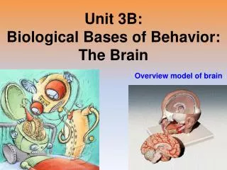
Brain & Cranial Nerves. Dr. Michael P. Gillespie. Major Parts of the Brain. Brain stem – continuous with the spinal cord. Medulla oblongata. Pons. Midbrain. Cerebellum – posterior to the brain stem. Major Parts of the Brain. Diencephalon – superior to the brain stem. Thalamus.

E N D
Brain & Cranial Nerves Dr. Michael P. Gillespie
Major Parts of the Brain • Brain stem – continuous with the spinal cord. • Medulla oblongata. • Pons. • Midbrain. • Cerebellum – posterior to the brain stem.
Major Parts of the Brain • Diencephalon – superior to the brain stem. • Thalamus. • Hypothalamus. • Cerebrum – supported on the diencephalon and brain stem. • Largest part of the brain.
Brain Blood Supply • Arteries • Internal carotid arteries • Vertebral arteries • Veins • Internal jugular veins
Brain Blood Flow • The brain consumes about 20% of the oxygen and glucose used at rest. • A brief slowing of blood flow may cause unconsciousness.
Brain Blood Flow • An interruption of blood flow for 1 to 2 minutes impairs neural function. • Total deprivation of oxygen for 4 minutes causes permanent injury. • If the blood entering the brain has a low level of glucose, mental confusion, dizziness, convulsions, and loss of consciousness may occur.
Blood Brain Barrier • The blood-brain barrier (BBB) protects the brain from harmful substances and pathogens. • It prevents the passage of many substances from the blood to the brain tissue. • Tight junctions seal together endothelial cells of brain capillaries. • Astrocytes selectively allow some substances through and not others.
Breaching the BBB • The BBB prevents the passage of harmful substances into the brain, but it also prevents the passage of useful drugs. • Drugs are injected in a concentrated sugar solution to facilitate passage. • The high osmotic pressure causes cells lining the barrier to shrink and makes the membrane “leaky”.
Protective Coverings • Cranial Meninges. • Dura mater. • Arachnoid mater. • Pia mater.
Cerebrospinal Fluid (CSF) • Clear colorless liquid. • Protects the brain and spinal cord from chemical and physical injuries. • Carries oxygen, glucose, and other needed chemicals from the blood to the neurons and neuroglia. • Circulates in the subarachnoid space (between the arachnoid mater and pia mater).
Protective Coverings • Extensions of the dura mater separate the parts of the brain. • Falx cerebri – separates the two hemispheres of the cerebrum. • Falx cerebelli – separates the two hemispheres of the cerebellum. • Tentorium cerebelli – separates the cerebrum from the cerebellum.
Formation of CSF in the Ventricles • CSF is formed in the ventricles. • Formed by ependymal cells that cover the choroid plexuses of the ventricles.
Formation of CSF in the Ventricles • There are 4 ventricles. • Functions of CSF. • Mechanical protection. • Shock absorption. • Buoys the brain. • Chemical protection – optimal chemical environment. • Circulation – medium of exchange for wastes and nutrients.
Hydrocephalus • Abnormalities of the brain can interfere with drainage of CSF from the ventricles and subarachnoid space. • CSF pressure increases causing hydrocephalus. • In infants this causes the fontanels to budge.
Hydrocephalus • Tumors, inflammation, developmental malformations can all cause hydrocephalus. • Pressure buildup can damage the delicate nervous tissue. • A surgeon can implant a drain line called a shunt to divert CSF. • In adults, hydrocephalus may occur after head injury, meningitis, or subarachnoid hemorrhage.
Brain Stem • Between the brain and spinal cord. • 3 regions. • Medulla oblongata. • Pons. • Midbrain.
Medulla Oblongata • A continuation of the spinal cord. • Sensory (ascending) tracts and motor (descending) tracts travel through the white matter of the medulla. • Many nerves decussate (cross over) in the medulla.
Medulla Oblongata • Cardiovascular center regulates the heartbeat and the diameter of the blood vessels.
Medulla Oblongata • The medullary rhythmicity area adjusts the rhythm of the breathing and controls reflexes for vomiting, coughing, and sneezing.
Medulla Oblongata • The nuclei for the following cranial nerves reside in the medulla: • VIII (vestibulocochlear). • IX (glossopharyngeal). • X (vagus). • XI (accessory). • XII (hypoglossal).
Pons • Pneumotaxic area and apneustic area regulate breathing. • Nuclei for cranial nerves V (trigeminal), VI (abducens), VII (facial), and VIII (vestibulocochlear).
Midbrain • The midbrain or mesencephalon contains the superior colliculi (visual actvities) and inferior colliculi (auditory pathways). • The midbrain contains the substantia nigra which release dopamine to help control subconscious muscle activities. Loss of these neurons results in Parkinson disease. • Cranial nerves III (oculomotor) and IV (trochlear) originate here.
Cerebellum • The second largest part of the brain. • A main function of the cerebellum is to evaluate how well movements are being carried out and correct for discrepancies. This helps to “smooth out” movements.
Diencephelon • Epithalamus. • Contains the pineal gland which secretes melatonin. • Thalamus. • Relays sensory information to the cortex. • Provides crude perception of touch, pressure, pain, and temperature.
Diencephelon • Subthalamus. • Controls body movements. • Hypothalamus. • Controls and integrates activities of the ANS. • Regulates emotional and behavioral patterns. • Regulates cicadian rhythms. • Regulates eating and drinking behavior. • Produces hormones oxytocin and ADH.
Cerebrum • Sensory areas interpret sensory impulses. • Motor areas control muscular movement. • Association areas function in emotional and intellectual processes. • Basal areas regulate gross muscle movements and regulate muscle tone. • Limbic system functions in survival behaviors.
Brain Injuries • Concussion – an abrupt, temporary loss of consciousness following a blow to the head. • Most common brain injury. • Signs – headache, drowsiness, lack of concentration, confusion, amnesia.
Brain Injuries • Contusion – bruising of the brain due to trauma and includes leakage of blood. • Signs - immediate loss of consciousness, transient cessation of respiration, decreased blood pressure.
Brain Injuries • Laceration – tear of the brain usually from a skull fracture or gunshot wound. • Rupture of large blood vessels. • Consequences – cerebral hematoma (localized pool of blood, usually clotted), edema, and increased intracranial pressure.
Cerebral Cortex Areas and Functions • Sensory areas – receive and interpret sensory information.
Cerebral Cortex Areas and Functions • Motor areas – initiate movements. • Association areas – deal with integrative functions: • Memory. • Emotions. • Reasoning. • Will. • Judgement. • Personality. • Intelligence.
Sensory Areas • Primary somatosensory area – receives sensations for touch, proprioception, pain, itching, tickle, and thermal sensations. • Located in the postcentral gyrus of the parietal lobes. • Primary visual area. • Primary auditory area. • Primary gustatory area – taste. • Primary olfactory area.
Motor Areas • Primary motor area – located in the precentral gyrus of the frontal lobe. • Broca’s speech area – coordinates the contractions of speech and breathing muscles.
Association Areas • Somatosensory association area – integrates and interprets sensations. • Visual association area – evaluates what is seen. • Auditory association area – evaluates sounds.
Association Areas • Wernicke’s (posterior language) area – interprets the meaning of speech. • Common integrative area. • Premotor area – controls learned skilled movements. • Frontal eye field area – controls voluntary scanning movements of the eyes.