Download
1 / 14
140 likes | 359 Views
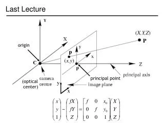
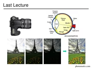
Vision. Last Lecture. Organization of the Visual System continued Blindsight What/Where pathways Their discovery, their neural retinal origins. This Lecture. What/Where pathways their manifestations in humans Examining the what pathway The Visual Agnosias.

E N D
Vision Last Lecture • Organization of the Visual System continued • Blindsight • What/Where pathways • Their discovery, their neural retinal origins
This Lecture • What/Where pathways • their manifestations in humans • Examining the what pathway • The Visual Agnosias
Single Units & Receptive Fields • Location(s) on sensory surface that causes cell firing (spatial r.f.). • Region of the retina from which a cell gets input. • Stimulus attributes that cause cell firing. • Motion • wavelength (color) • shape • Specialized modules monitoring the world for the attributes that “turn them on” • consist of cells with specific receptive field properties
What vs. Where in Humans • Focal lesions produce selective color or motion blindness. • cortical color blindness (achromatopsia) • motion blindness (akinetopsia) • Positron Emission Tomography (PET) studies in Humans reveal: • Motion Area distinct from color area • (e.g. Zeki and colleagues, 1990.)
Achromatopsia • Inability to perceive color-- color knowledge and color naming can be intact. • acquired after cortical brain damage • distinct from congenital color blindness (photoreceptor abnormality) • Subjective reports... • Lesion locus: bilateral occipito-temporal lesions associated w/upper visual field deficits.
Akinetopsia: Motion blindness due to focal brain damage (Zihl and colleagues, 1983) Characteristics of this famous case: • no blindness as assessed by perimetry • mild calculation and speech difficulties • all other aspects of vision are within normal range • subjective reports... • Lesion locus: input to dorsal pathway (V5/MT)
PET Activation Study Logic • Perceptual/cognitive activity mediated by localized neural activity. • Blood flow changes accompany regional changes in neural activity. • Blood flow changes measured by monitoring distribution of radioactive tracer.
Regions active in response to COLOR Regions active in response to MOTION
A Schematic of What vs. Where AKINETOPSIA ACRHOMOTOPSIA
Attribute-specific deficits • Reveal unique functions performed by distinct areas. • Special purpose modules perform operations in parallel. • Functions of many modules still undefined. • Consider deficits of form, object recognition...