Download
1 / 25
280 likes | 735 Views
Two -methods are generally used, (wet method) and (dry and fix method (. for Studying Microbes with a Compound Microscope. Wet Method . There are two primary methods generally used for studying microorganisms in wet conditions wet mount method hanging drop method. The wet mount.

E N D
Two -methods are generally used, (wet method) and (dry and fix method (.for Studying Microbes with a Compound Microscope • Wet Method.There are two primary methods generally used for studying microorganisms in wet conditions • wet mount method • hanging drop method.
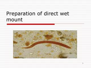
The wet mount A drop of fluid containing microorganisms. The fluid spreads out in a thin layer between coverslip and slide.
The hanging Drop Method It is used to observe the motility, germination, or fission of microorganisms.
Dry and fix method Microorganisms, particularly bacteria, being too small need their permanent preparation be made by drying and fixing them on clean slide with or without staining.
For preparing a dry mount: A drop of distilled water with a small amount of culture is spread as a thin smear on a clean slide. The smear is allowed to dry and it is then 'fixed' by passing it through a flame two to three times with the smeared slide away from the flame. If desired, this dried and fixed amount may be stained and the preparation dried again for observation under the microscope.
Bacterial Shapes : Bacteria have three basic shapes: Round cells (Cocci) , Rod-shaped cells are bacilli , spiral-shaped cells
Types of stain • Chromophore • Positive: basic stain • Attaches to cell wall and bacterium appears colored • Direct Stain • Negative: acidic stain • Repelled from cell wall; stains background • Negative Stain • Simple stains • To observe basic external structures of cell with brightfield scope (cellular morphology) • Reagent: Methylene Blue
Smears • Heat Fixing • Kill • Stops autolysis • Adherence to slide • Air dry first to prevent lysis (boiling)
Use of a single basic dye is called a simple stain. A mordant may be used to hold the stain or coat the specimen to enlarge it.
The Gram stain classifies bacteria into gram-positive and gram-negative. Gram-positive bacteria tend to be killed by penicillin and detergents. Gram-negative bacteria are more resistant to antibiotics.
Gram Stain • Purpose • To view cellular morphology • Diagnostic purposes: What type of cell wall a bacterium has • Gram Positive • Thicker peptidoglycan layer and no outer membrane • Gram Negative • Thinner Peptidoglycan and an out membrane
Reagents for Gram Stain • Crystal Violet (purple) • Primary stain; positive stain • Stains cell wall purple • Iodine • Mordant • Combines with CV to form an insoluble complex that gets trapped in thicker peptidoglycan layers • What happens if you skip this step? • Ethanol • Decolorizer • CV-I complex washed out of Gram negative organisms because it cannot be trapped by peptidoglycan layer; flows right through outer membrane • Safranin (pink) • Counterstain • Simple positive stain that provides contrasting dye for decolorized cells (Gram negative) • Stains all cells, but only the negative ones actually appear pink.
The characteristic compound found in all true bacterial cell walls is peptidoglycan. The amount of PPG is among one of the differences between the GP and GN cell walls. Gram-positive cell walls Gram-negative cell walls Thick peptidoglycan 90% peptidoglycan Teichoic acids 1 layer Not many polysaccharides In acid-fast cells, contains mycolic acid Thin peptidoglycan 5-10% peptidoglycan No teichoic acids 3 layers Outer membrane has lipids, polysaccharides No acid- fast cells (mycolic acid)
The crucial step in the staining process is the decolorizing step. The most accepted theory about the rationale for the Gram staining process is the one proposed by Salton. This theory relies on the fact that the PPG is found in layers and the stain molecules are trapped within the many layers of the GP CW when they form the complex with the mordant Iodine molecules. Since the GN CWs lack much PPG the amount of stain captured in those CWs is much lesser. When the cells are treated with the decolorizer – the ethanol – this causes denaturation of the proteins in the outer membrane of the GN CWs resulting in gaping holes in these CWs that lead to the removal of the crystal violet-iodine complexes easily, leaving these cells unstained. The counterstain -safranin- thus is used to make these cells visible.
There are 4 conditions to be followed for a valid Gram staining procedure: Young cultures - must be young within 18-24hrs old (older cultures lose their Gram staining properties due to changes in the CWs as the cells get older) Thin smear thicker or uneven smears will result in uneven staining and decolorization Fresh reagents - of proper strength Control cultures - for a known GP bacterium and GN culture (S.aureus & E.coli)