Download
1 / 80
1.01k likes | 2.63k Views
Mechanical Ventilation: The Basics and Beyond. Presented By: Diana Gedamke, BSN, RN, CCRN Marion College - Fond du Lac. Mechanical Ventilation.

E N D
Mechanical Ventilation: The Basics and Beyond Presented By: Diana Gedamke, BSN, RN, CCRN Marion College - Fond du Lac
Mechanical Ventilation “…an opening must be attempted in the trunk of the trachea, into which a tube of reed or cane should be put; you will then blow into this, so that the lung may rise again…and the heart becomes strong…” Andreas Vesalius (1555)
Mechanical Ventilation • First described in 1555 • First used in patient care during the polio epidemic of 1955 • Medical students became human ventilators • First positive pressure ventilator was used at Massachusetts General Hospital
Indications for MV • Acute Respiratory Failure (66%) • Coma (15%) • Acute Exacerbation of COPD (13%) • Neuromuscular Disorders (5%) Esteban et al. How is mechanical ventilation employed in the intensive care unit? An international utilization review. Am J Respir Crit Care Med 2000;161: 1450 - 8.
Indications for MV • Acute Respiratory Failure • ARDS • Heart Failure • Pneumonia • Sepsis • Complications of Surgery • Trauma
Objectives of MV • Decrease WOB • Reverse life-threatening hypoxemia or acute progressive respiratory acidosis • Protect against ventilator-induced lung injury • Wean and extubate as soon as possible
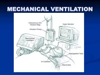
Caring for the Mechanically Ventilated Patient • Endotracheal tube • Position • Stability • Cuff inflation • Patency • Oral cavity • Trauma to lip and palate • Secretions
Caring for the Mechanically Ventilated Patient • Patient Position • Patient-Ventilator Synchrony • Physical Assessment • VS, General assessment, breath sounds • Ventilator Mode • Alarms • Reason for intubation • MV in disease states • Does patient still need to be intubated and mechanically ventilated?
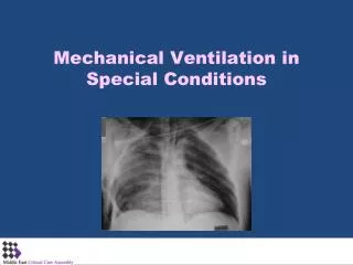
Nasal or oral Size (6 – 8.5 cm) Position 21 cm from the teeth in women & 23 cm in men Confirm position by CXR (even when breath sounds are bilateral) 3 - 5 cm above carina Affects of head position Endotracheal Tube
Endotracheal Tube • Position • End Tidal CO2 to confirm ETT placement
Complications of Intubation and Mechanical Ventilation • Sinusitis - occurs in > 25% of patients ventilated > 5days; nasal > oral; subtle findings (unexplained fever & leukocytosis), polymicrobial • Laryngeal damage - ulceration, granulomas, vocal cord paresis, laryngeal edema • Aspiration/Ventilator-associated pneumonia - occurs despite cuffed tube • Tracheal necrosis - tracheal stenosis, tracheomalacia • Death – ventilator malfunction/inadvertent disconnection/endotracheal tube dysfunction/VAP
Preventing Infection • Sterile suctioning • assess as routine part of assessment; only suction when patient needs it • Elevate HOB to 45 degrees
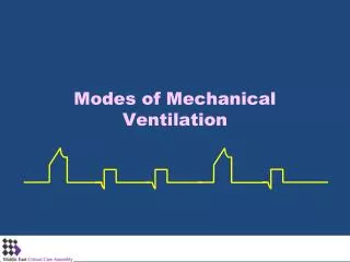
Positive Pressure Ventilation • Volume-cycled ventilation • Pressure-preset ventilation
Volume-cycled Ventilation • Delivers a preset volume of gas with each machine breath—airway pressures increase in response to the delivered breath • Airway pressures are higher in patients with low compliance or high resistance—high pressures indicate risk of ventilator-induced lung injury
Volume-cycled Ventilation • Assist Control (AC) • Synchronized Intermittent Mandatory Ventilation (SIMV)
Assist-control (AC) • Most widely used mode of MV • Delivers a minimum number of fixed-volume breaths • Patients can initiate extra assisted breaths (will get full set volume with each effort)
Synchronized Intermittent Mandatory Ventilation (SIMV) • Delivers preset number of fixed-volume breaths • Patient can breathe spontaneously between breaths (rate and depth determined by patient) • Patients often have trouble adapting to intermittent nature of ventilatory assistance
Pressure-preset Ventilation • Delivers a predefined target pressure to the airway during inspiration • Resulting tidal volume (VT) and inspiratory flow profile vary with the impedance of the respiratory system and the strength of the patient’s inspiratory efforts • Includes pressure-control (PC) and Pressure support (PS)
Pressure-control (PC) Ventilation • Delivers a preset gas pressure to the airway for a set time and at a guaranteed minimum rate • Patient can breathe in excess of set rate • Tidal volume achieved depends on pressure level, lung mechanics, and patient effort • Inspiratory flow rate variable
Pressure Support (PS) • Delivers preset airway pressure for each breath • Variable parameters: Inspiratory and expiratory times (respiratory rate), flow rate, and tidal volume (VT)
SIMV + PS • Spontaneous breaths allowed in SIMV are assisted by PS
New Modes of MV • New modes often introduced • Involves nothing more than a modification of the manner in which positive pressure is delivered to the airway and of the interplay b/n mechanical assistance and patient’s resp effort • Goals: enhance respiratory muscle rest, prevent deconditioning, improve gas exchange, prevent lung damage, improve synchrony, foster lung healing
Ventilator Settings • Respiratory rate • Tidal volume • FiO2 • Inspiratory:Expiratory (I:E) ratio • Pressure limit • Flow rate • Sensitivity/trigger • Flow waveform
Inspiratory Flow (V) Waveform Square waveform Decelerating Waveform (constant flow) (decelerating flow)
Inspiratory Flow (V) Waveform • Square waveform: volume of gas is evenly distributed across inspiratory time. Has highest peak pressure and lowest mean airway pressure. Ideal for those at risk for autopeeping due to short inspiration time
Inspiratory Flow (V) Waveform • Decelerating waveform: Volume of gas flow is high at the beginning of inspiration then tapers off toward the end of the breath. Has lowest peak pressure and highest mean airway pressure. Increased inspiratory time; useful in ARDS.
Patient-Ventilator Synchrony • Check for: • Symmetric chest inflation • Regular breathing pattern • Respiratory rate < 30 bpm • Synchrony between patient effort and machine breath • Paradoxical breathing
Patient-Ventilator Synchrony • Inspiratory effort expended by patients with acute respiratory failure is 4 - 6 x normal • Don’t eliminate respiratory effort: causes deconditioning and atrophy
Patient-Ventilator Asynchrony • Possible causes: • Anxiety or pain • Ventilator settings may not be appropriate: check ABG and alert individual responsible for ventilator orders • Auto-PEEP • Pneumothorax
Definitions • PEEP – positive-end-expiratory pressure applied during mechanical ventilation • CPAP - continuous positive airway pressure applied during spontaneous breathing
PEEP • Improves oxygenation - increases functional residual capacity (FRC) above closing volume to prevent alveolar collapse • permits reduction in FIO2 • Reduces work of breathing • Increases intrathoracic pressure - decreases venous return to right heart - decreases CO • Titrate to least amt. necessary to achieve O2 sat > 90% or PO2 > 60 mm Hg with FiO2 < 0.6
Auto-PEEP • Auto-PEEP/intrinsic PEEP (PEEPi)/inadvertent PEEP/occult PEEP - positive end expiratory alveolar pressure occurring in the absence of set PEEP. Occurs when expiratory time is inadequate.
Definitions • Peak Airway Pressure (Ppk) • An increase in Ppk indicates either an increase in airway resistance or a decrease in compliance (or both). • Plateau Pressure (Ppl) - end-inspiratory alveolar pressure
Ventilator-Induced Lung Injury • High volumes and pressures can injure the lung, causing increased permeability pulmonary edema in the uninjured lung and enhanced edema in the injured lung • Alveolar overdistention + repeated collapse and re-opening of alveoli
Mechanical Ventilation in Obstructive Lung Disease • resistance to expired flow • results in air trapping/hyperinflation • hyperinflation may result in cardiopulmonary compromise • Goal: meet minimal requirements for gas exchange while minimizing hyperinflation • Allow increased time for expiratory flow
Increasing Time for Exhalation • Decrease inspiratory time • Increase flow rate • Square waveform • Decrease minute ventilation (VE) • RR x TV
Monitoring Patients with Obstructive Lung Disease Requiring Mechanical Ventilation • Monitor plateau pressure: in general, Pplat < 30 cm H20 to decrease risk of hyperinflation and alveolar overdistension • Permissive hypercapnia
Respiratory Failure Due to Asthma • Watch for overventilation post intubation • High Ppk common • May require sedation to establish synchronous breathing with ventilator • Avoid paralytics • Ventilate as stated above (Increase exhalation time by decreasing RR and TV, increasing inspiratory flow rate, and using square waveform) • May want to use SIMV
Respiratory Failure Due to COPD • Ppk typically not as elevated as in asthma; when it is, think other pathologic processes • Many patients with COPD have chronic hypercapnia; ventilatory support titrated to normalize pH and not PCO2 • Small levels of set PEEP may decrease WOB • May try NIPPV
Noninvasive Positive Pressure Ventilation (NIPPV) • Cooperative patient • Functionally intact upper airway • Minimal amount of secretions • Done by full face or nasal mask • Watch for gastric distension; may increase risk of aspiration • May use standard ventilators • Monitor patients closely for decompensation and need for intubation