Download
1 / 9
100 likes | 269 Views
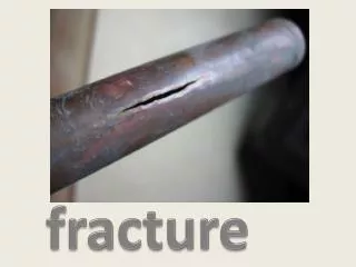
Fracture Descriptions. Dr. LeeAnn Pack Dipl. ACVR. Can you describe this fx?. Answer. Transverse to short oblique (simple) complete fxs are seen in the distal 1/3 of the diaphysis of the radius and ulna The distal fragments are displaced lateral and cranial.

E N D
Fracture Descriptions Dr. LeeAnn Pack Dipl. ACVR
Answer • Transverse to short oblique (simple) complete fxs are seen in the distal 1/3 of the diaphysis of the radius and ulna • The distal fragments are displaced lateral and cranial. • Mild soft tissue enlargement is present in the region of the fx • The fx appears closed radiographically
Answer • A long oblique to spiral complete fx is seen in the mid diaphysis of the tibia • The distal fragment is displaced lateral, is slightly overriding on the CC view and slight cranial displacement is also noted. • Small fissures can be seen extending into the distal fragment • Mild soft tissue enlargement is present in the region of the fx • There is also a rotational mal alignment present as the stifle is CC and the tarsus is oblique medially
Answer continued • A segmental fracture is seen in the proximal to mid 1/3 of the fibula. • The distal fragment is displaced minimally and the segmental portion is displaced laterally
Answer • A long oblique complete fx is seen in the mid diaphysis of the tibia • The distal fragment is displaced cranially and is slightly overriding • A single large cortical fragment is also seen cranial and slightly proximal to the main fx site • Mild soft tissue enlargement is seen in the region of the fx
Answer continued • A comminuted fx of the calcaneus is also seen – there are multiple (~3 fragments) present but they are minimally displaced. • The fractures appear closed radiographically • A CC view of the limb is is also needed