Download

1 / 7
160 likes | 839 Views
Gram Staining Discussion. May 27, 2011. Let’s Practice!. Use the sheets given to you and complete a Gram stain on the bacteria cultures in the petri dishes Use what we have already mentioned in class to determine which bacteria are Gram positive and which are Gram negative .

E N D
Gram Staining Discussion May 27, 2011
Let’s Practice! • Use the sheets given to you and complete a Gram stain on the bacteria cultures in the petri dishes • Use what we have already mentioned in class to determine which bacteria are Gram positive and which are Gram negative
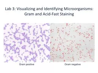
Types of Gram Stains • Gram positive • Bacteria cells containing peptidoglycan absorb only the violet dye • Appear purple in the microscope
Types of Gram Stains (cont.) • Gram negative • Bacteria with a second (outer) layer of lipid & carbohydrate molecules • Extra layer allows purple dye to be removed during decolorizing stage and red dye to be absorbed • Appear pink in the microscope
Why is there a difference? • Thick peptidoglycan of the Gram + retains the purple dye even after decolorizing step
Why do we use gram staining? • Almost always the first step in the identification of a bacterial organism • Allows a doctor to prescribe treatment with an appropriate antibiotic while waiting for more specific tests, such as a culture, to be completed • Not all bacteria can be definitively classified by this technique
Steps of Gram Staining • Place a slide with a BACTERIAL smear • STAIN the slide with crystal violet for 1-2 min. • Pour off the stain • Flood slide with Gram's IODINE for 1-2 min. • Pour off the iodine • DECOLORIZE by washing slide with acetone • WASH slide with water to remove the acetone • Flood slide with safranin COUNTERSTAIN for 2 min. • WASH with water • Blot excess water and DRY in hand Gram Stain Animation