Download
1 / 59
630 likes | 2.03k Views
Plastic and reconstructive surgery. Dr Yaseen Abdullah Plastic surgeon. Learning objectives To understand The spectrum of plastic surgical techniques used to restore bodily form and function The relevant anatomy and physiology of tissues used in reconstruction

E N D
Plastic and reconstructive surgery Dr Yaseen Abdullah Plastic surgeon
Learning objectives To understand • The spectrum of plastic surgical techniques used to restore bodily form and function • The relevant anatomy and physiology of tissues used in reconstruction • The various skin grafts and how to use them appropriately • The principles and use of flaps
Plastic : Greek word : to mould or shape • Plastic and reconstructive surgery involve using various techniques to restore form and function to the body when tissues have been damaged by injury , cancer, or congenital loss
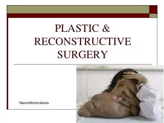
The scope of plastic surgery The tools of reconstruction are used for a wide range of conditions: ●● trauma: ●● soft-tissue loss (skin, tendons, nerves, muscle); ●● hand and lower limb injury; ●● faciomaxillary; ●● burns; ●● cancer: ●● skin, head and neck, breast, soft tissue sarcoma; ●● congenital: ●● clefts and craniofacial malformations; ●● skin, giant naevi, vascular malformations; ●● urogenital; ●● hand and limb malformations; ●● miscellaneous: ●● Bell’s (facial) palsy; ●● pressure sores; ●● aesthetic surgery; ●● chest wall reconstruction
Classification of wound closure and healing ●● Primary intention Wound edges opposed Normal healing Minimal scar ●● Secondary intention Wound left open Heals by granulation, contraction and epithelialisation Increased inflammation and proliferation Poor scar ●● Tertiary intention (also called delayed primary intention) Wound initially left open Edges later opposed when healing conditions favorable
Skin grafts • a surgical operation in which a piece of healthy skin is transplanted to a new site on a patient's body. • Transferred without their blood supply • Skin grafts can be: • A split-thickness skin graft (STSG) is a skin graft including the epidermis and part of the dermis. • A full-thickness skin graft (FTSG)consists of the epidermis and the entire thickness of the dermis.
Thicker knife-gap settings give rise to fewer but briskerbleeding points on the donor site
Meshed grafts have more size and less haematoma riskbut they are ugly with more contracture
Split-thickness skin grafts ●● Thicker knife-gap settings give rise to fewer but brisker bleeding points on the donor site. ●● Thicker grafts heal with less contracture and are more durable. ●● Thinner donor sites heal better. ●● Grafts are hairless and do not sweat (these structures are not transferred).
How does a skin graft survive? • imbibition of plasma from the wound bed intially • after 48 hours, fine anastomotic connections are made, which lead to inosculation of blood. • Capillary ingrowththen completes the healing process with fibroblast maturation. Because only tissues that produce granulation will support a graft, it is usually contraindicated to use grafts to cover exposed tendons, cartilage or cortical bone.
Causes of graft failure • Pus • Haematoma • Exudate • Dead or non vascularized bed • Shearing force • group A β-haemolyticStreptococcus can destroy split grafts completely (and also convert a donor site to a full-thickness defect
Skin flaps • A skin flap consists of skin and subcutaneous tissue that survives based on its own blood supply. • Transferred with their blood supply
Classification of flaps according to blood supply • Random flaps have no named blood supply • Axial flaps are supplied by a named artery and vein. This allows for a larger area to be freed from surrounding and underlying tissue, leaving only a small pedicle containing the vessels. • Pedicled flaps remain attached to the donor site via a pedicle that contains the blood supply • Island flap attached only to the blood vessel • free flap: Fully detached from vascular supply and reconnected to recipient vessels using microvascular technique
Classification of flaps according to location a. Local flap: Shares side with the defect b. Regional flap: In same region of the body as the defect, but does not share defect margin. c. Distant flap: Not in the region of the defect, located in a different part of the body
Classification of flaps according to Method of transfer a. Advancement b. Transposition c. Rotation
Eg. Local (transposition, random)flap: share border with the defect
Tissue expansion A technique that involves placing a device – usually an expandable balloon constructed from silicone – beneath the tissue to be expanded, and progressively enlarging the volume with fluid while the overlying tissue accommodates to the changed vascular pressure