Download
1 / 71
740 likes | 1.14k Views
Muscular System: Histology and Physiology. Chapter 9. Muscular System Functions. Body movement Maintenance of posture Respiration Production of body heat Communication Constriction of organs and vessels Heart beat. Criteria for Naming Muscles. Shape : romboideus, trapezius, biceps

E N D
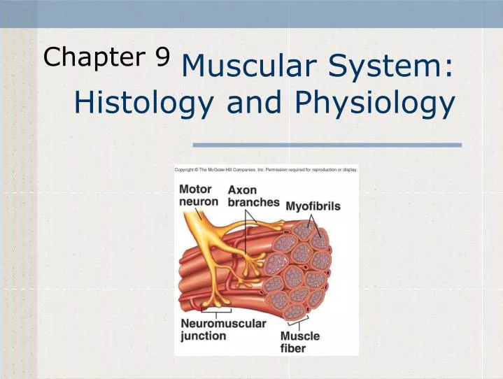
Muscular System:Histology and Physiology Chapter 9
Muscular System Functions • Body movement • Maintenance of posture • Respiration • Production of body heat • Communication • Constriction of organs and vessels • Heart beat
Criteria for Naming Muscles • Shape: romboideus, trapezius, biceps • Location: pectoralis (chest) intercostal (ribs) • Attachment: zygomaticus, sternocleidomastoid • Size: maximus, minimus, brevis, longis • Orientation of fibers: rectus (straight), oblique (slanting) • Relative position (lateral, medial, internal, external) • Function: adductor, flexor, extensor, pronator
Properties of Muscle • Contractility • Ability of a muscle to shorten with force • Excitability • Capacity of muscle to respond to a stimulus • Extensibility • Muscle can be stretched to its normal resting length and beyond to a limited degree • Elasticity • Ability of muscle to recoil to original resting length after stretched
Skeletal Muscle Cardiac Muscle Smooth Muscle
Skeletal Muscle Structure • Muscle fibers or cells • Develop from myoblasts • Numbers remain constant • Hypertrophy – increase in the size of each fiber. • Connective tissue • Nerve and blood vessels
Connective Tissue, Nerve, Blood Vessels • Connective tissue • External lamina • Endomysium • Perimysium • Fasciculus • Epimysium • Fascia • Binds adjacent muscles or overlying skin. • Nerve and blood vessels • Abundant
Sliding Filament Model • Actin myofilaments sliding over myosin to shorten sarcomeres • Actin and myosin do not change length • Shortening sarcomeres responsible for skeletal muscle contraction • During relaxation, sarcomeres lengthen
Physiology of Skeletal Muscle • Nervous system • Controls muscle contractions through action potentials • Resting membrane potentials • Membrane voltage difference across membranes (polarized) • Inside cell more negative and more K+ • Outside cell more positive and more Na+ • Must exist for action potential to occur
Ion Channels • Types • Ligand-gated • Example: neurotransmitters • Voltage-gated • Open and close in response to small voltage changes across plasma membrane
Action Potentials • Phases • Depolarization • Inside plasma membrane becomes less negative • Repolarization • Return of resting membrane potential • All-or-none principle • Like camera flash system • Propagate • Spread from one location to another • Frequency • Number of action potential produced per unit of time
Neuromuscular Junction • Synapse or NMJ • Presynaptic terminal • Synaptic cleft • Postsynaptic membrane or motor end-plate • Synaptic vesicles • Acetylcholine: Neurotransmitter • Acetylcholinesterase: A degrading enzyme in synaptic cleft
Excitation-Contraction Coupling • Mechanism by which an action potential causes muscle fiber contraction • Involves • Sarcolemma • Transverse or T tubules • Terminal cisternae • Sarcoplasmic reticulum • Ca2+ • Troponin
Muscle Twitch • Muscle contraction in response to a stimulus that causes action potential in one or more muscle fibers • Phases • Lag or latent • Contraction • Relaxation
Stimulus Strength and Muscle Contraction • All-or-none law for muscle fibers • A motor unit contracts with a consistent force in response to each action potential • Sub-threshold stimulus • Threshold stimulus • Stronger than threshold • Motor units • Single motor neuron and all muscle fibers that it innervates • Graded for whole muscles • Strength of contractions range from weak to strong depending on stimulus strength
Multiple Motor Unit Summation • A whole muscle contracts with a small or large force depending on number of motor units stimulated to contract • Muscle performing delicate and precise movements have motor units with smaller numbers of fibers
Multiple-Wave Summation • As frequency of action potentials increase, frequency of contraction increases • Incomplete tetanus • Muscle fibers partially relax between contraction • Complete tetanus • No relaxation between contractions • Multiple-wave summation • Muscle tension increases as contraction frequencies increase • Due to increased calcium concentration around myofibrils and more complete stretching of muscle elastic elements
Treppe • Increase in the force of contraction during the first few contractions of a rested muscle. • Occurs in muscle rested for prolonged period • Each subsequent contraction is stronger than previous until all equal after few stimuli • Due to Ca++ ion levels around myofibrils and increased temperature of muscle • Enzymes for muscle contraction respond more effectively at higher temperature.
Types of Muscle Contractions • Isometric: No change in length but tension increases • Postural muscles of body • Isotonic: Change in length but tension constant • Concentric: Overcomes opposing resistance and muscle shortens • Eccentric: Tension maintained but muscle lengthens • Muscle tone: Constant tension by muscles for long periods of time
Fatigue • Decreased capacity to work and reduced efficiency of performance • Usually follows a period of activity • Types • Psychological (in CNS) • Depends on emotional state of individual • Perception that muscle is too tired ( • Home court advantage • Muscular • Results from ATP depletion in muscle • Synaptic • Occurs in NMJ due to lack of acetylcholine
Energy Sources • ATP provides immediate energy for muscle contractions from 3 sources • Creatine phosphate • During resting conditions stores energy to synthesize ATP • Exhausted quickly (10-15 sec.) • Anaerobic respiration • Occurs in absence of oxygen and results in breakdown of glucose to yield ATP and lactic acid • Aerobic respiration • Requires oxygen and breaks down glucose to produce ATP, carbon dioxide and water • More efficient than anaerobic • Oxygen Debt • After anaerobic respiration, aerobic respiration is higher than normal to replace creatine phosphate and convert lactic acid to glucose.
Slow and Fast Fibers • Slow-twitch or high-oxidative • Contract more slowly, smaller in diameter, well developed blood supply, more mitochondria and high myoglobin content, more fatigue-resistant than fast-twitch • Fast-twitch or low-oxidative • Respond rapidly to nervous stimulation, less blood supply, fewer and smaller mitochondria, lower myoglobin content than slow-twitch, fatigue easily. • Two types: • Fast twitch fatigable fibers • Fast twitch fatigue resistant (highly trained muscle) • Distribution of fast-twitch and slow twitch • Most muscles have both but varies for each muscle