Download
1 / 77
800 likes | 1k Views
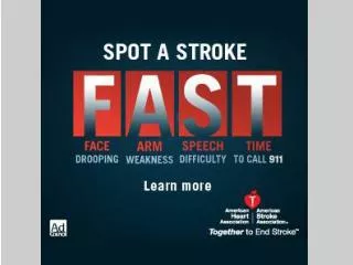
Neurosensory: Stroke and Brain Tumors. Part #1 Stroke (Brain attack/CVA). A. Pathophysiology/etiology Normal brain physiology and stroke. Ranks 3 rd as cause death Blood supply to one hemisphere is typically blocked, hence terms right & left stroke

E N D
Neurosensory: Stroke and Brain Tumors Part #1 Stroke (Brain attack/CVA)
A. Pathophysiology/etiology Normal brain physiology and stroke • Ranks 3rd as cause death • Blood supply to one hemisphere is typically blocked, hence terms right & left stroke • Functioning brain depends on continuous blood supply for oxygen and glucose & remove end products metabolism
Hypertension Heart disease Atherosclerosis Diabetes mellitus Medications: birth control pills, substance abuse- cocaine, heroin Sedentary life style Obesity High cholesterol diet Smoking Stress Age > 65 yrs Sickle cell disease Risk factors for stroke
Brain dysfunction & length of time without blood supply • Brain function depends on collateral circulation and amount of cerebral edema • TIA- neuro deficits last < 24 hrs • RIND- neuro deficits last > 24 hrs but reverse not greater than 21 days • CVA- irreversible brain damage with residual neuro deficits • Stroke-in-evolution- progressive neuro deficits developing over hours or days. Usual cause thrombosis
Ischemic stroke Occlusion of artery Generally do not lose consciousness Better prognosis than hemorrhagic May have TIA’s before Thrombosis or embolism Hemorrhagic stroke Bleed occurs with activity Usually rapid onset Generally loss of consciousness Poorer prognosis Intracranial or subarachnoid Disease process
Ischemic stroke Thrombosis • Most common cause of a stroke • Cause- narrowing of artery from atherosclerotic plaques • Blood is blocked to part of brain that the artery supplies • Often occurs in older individuals who are at rest/sleeping • Tend to form in large arteries that bifurcate, internal carotid artery common site • Can begin as TIA’s, present as stroke-in-evolution, or have completed stroke outright
Ischemic stroke Embolism • Caused by: clotted blood from other arteries in the body (heart during atrial fibrillation) fat, bacteria (endocarditis) or air • Emboli circulate until reach an artery in brain that is too narrow to pass through • Usually awake with rapid onset • Extent damage is less severe and recovery faster than other strokes
Hemorrhagic stroke Intracranial hemorrhage (ICH) • Caused by ruptured artery in the brain • Bleeding varies in size from petechial to massive, edema occurs around the bleed • Blood may form hematoma or be diffuse within the brain • Usually occurs rapidly with the deep arteries • Hypertension is main cause • Most common cause of death due to a stroke • Have more extensive residual deficits and slower recovery than other causes of stroke
Hemorrhagic Subarchnoid hemorrhage (SAH) • Caused by bleeding into subarchnoid space from • Extension of a intracranial hemorrhage • Aneurysm • AV malformation
B. Common manifestations/complications- by body systems
By artery affected by occlusion or hemorrhage Internal carotid
Middle cerebral artery • Contralateral motor loss in the arm and the lower part of the face (central facial palsy) • Contralateral sensory loss in face and arm • Homonymous hemianopsia • Left middle-communication deficits • Right- spatial/perceptual
Vertebral artery • Pain or numbness of involved side • Vertigo • Contralateral ataxia • Dysphagia, dysarthria • Cranial nerve dysfunctions
Motor deficits • Motor nerve pathways cross in the medulla (brainstem) Prefix hem- used to describe • Amount of motor involvement varies from weakness (-paresis) to paralysis (-plegia). • End paralysis can be flaccid or spastic depending on amount of damage to the motor strip • Initially flaccid and if progress spastic in 6-8 weeks.
Motor deficits • Characteristic body posture
Motor deficits • Facial palsy- (central/UMN) where lower part face affected • Bells palsy (LMN- 7th CN) where the whole side of face affected
Elimination Deficits • Partial loss of sensation (hemi) can affect perception of need to eliminate bowel/bladder • Cognitive problems may affect the social aspect of elimination • Level of consciousness, immobility, dehydration, diet changes
Sensory-perceptual deficits Lack of sensation/propriocetion • Lack of sensation (hemi)- inability to perceive/interpret pain, touch, pressure( post central gyrus) • Lack of/decrease in proprioception or the inability to know where body part is without having to look at it; body’s ‘position sense’
Sensory-perceptual deficits Visual field deficits • Disruption anywhere along the pathway • Homonymous hemianopsia- most common. Loss of half of visual field in each eye. Can’t see toward the same side as the paralysis
Inability of the senses to perceive stimuli that were previously familiar. May be any of the senses and varying degrees Inability to carry out purposeful task in the absence of paralysis or the individual carries out task inappropriately Sensory-perceptual deficits: Agnosia Apraxia
Sensory-perceptual deficits Neglect syndrome (unilateral neglect) • Attention disorder in which individual ignores affected part of the body, • Cannot integrate or use perceptions from affected side • More common in right CVA’s
Communication Deficits • Motor, speech, language, memory, reasoning, emotions can be affected • Dominant hemisphere for the brain centers is left in most individuals • Global (mixed) aphasia- both expressive and receptive aphasia • Dysarthria- difficulty with articulation or muscular control for speech. Sound like have mashed potatoes in their mouth
Communication Deficits Broca’s and Wernicke’s aphasia • Broca’s, expressive or nonfluent aphasia where unable to express- understands • Wernicke’s, receptive, fluent aphasia where unable to understand
Communication Deficits Normal process recovery • Begin with one word speech- swearing, ‘ouch’ • Progress to sayings – days of week, social speech, singing • Volitional- normal speech • Recovery may stop at any point
Cognitive and behavioral deficits • Change level consciousness- confusion to coma • Emotional liability • Loss of self control, decrease tolerance for stress • Intellectual changes resulting in memory loss, decreased attention span, poor judgment, inability to think abstractly
C. Therapeutic interventions Diagnostic tests • CT/MRI- bleeding, edema, tissue necrosis, shifting intracranial contents • Arteriogram- abnormal structures; vasospasm, stemosis • PET- cerebral blood flow and metabolic activity • Transcranial ultrasound doppler velocity of blood flow, degree of occlusion • Lumbar puncture- obtain CSF, bleeding
Therapeutic interventions Rehabilitation • Outpatient or in-house • Physical therapy • Occupational therapy • Speech therapy • Cognitive therapy
Therapeutic interventions Thrombolitic stroke • Medication • Thrombolitic agents to dissolve clot- 3 hrs!!! • Anticoagulants to prevent further extension • Antithrombolitic inhibit platelet phase of clot formation • Anticonvulsants • Surgical • Endarterectomy • Angioplasty, carotid artery stenting • Bypass superficial temporal to middle cerebral
Therapeutic interventions Embolic/intracranial stroke • Embolic stroke • Medications: If blood clot- anticoagulants, thrombolitic agents, antiarrhythmics; If bacterial- antibiotics • Intracranial hemorrhage (ICH) stroke • Bedrest • Medication- antihypertensives to normal BP • Surgery- remove hematoma if possible
D. Nursing assessment specific to stroke Health history & physical exam • Health history- • Risk factors; when symptoms began; describe symptoms; current medications (legal/illegal); other health problems • Physical exam- • Vital signs; neuro vital signs (LOC, pupils, motor, sensory); continued next slides
Nsg assess- neuro deficits common in stroke Motor • Movement, strength (with & without resistance), symmetry of all extremities • Pronator drift- detects weakness of upper extremity. Hold arms, palms up in front with eyes closed- should be able to hold for 30 seconds. Weakness pronates and drifts downward • Use similar techniques used to assess motor SCI- motor pathways affected begin motor strip brain • Test facial movement- smile/frown test for Bell’s (7th CN) and central facial (motor strip)
Nursing assess- neuro deficits common stroke Motor • EOM’s- head still, follow your finger in all quadrants. Eyes should move together (conjugate gauze) Abnormal: dysconjugate gauze; nystagmus; 3rd nerve palsy (occulomotor); 6th nerve palsy (abducens)
Nursing assess neuro deficits: Motor 3rd nerve palsy 6th nerve palsy
Nursing assess- neuro deficits common stroke Motor • Assess ability to void and move bowels • Assess communication ability • Assess cognitive and behavioral aspects
Nursing assess-neuro deficits common stroke Sensory deficits • Superficial sensation • With paperclip and eyes closed alternate sharp and dull ends • Reference is the sensory strip on the parietal side
Nursing assess- neuro deficits common strokeSensory- visual field loss common- homonymous hemianopia • Patients’ head in still position & cover one eye- test one at time • Move your wiggling finger into the patients field of vision- in all 6 quadrants • State when 1st sees