Download
1 / 17
170 likes | 172 Views
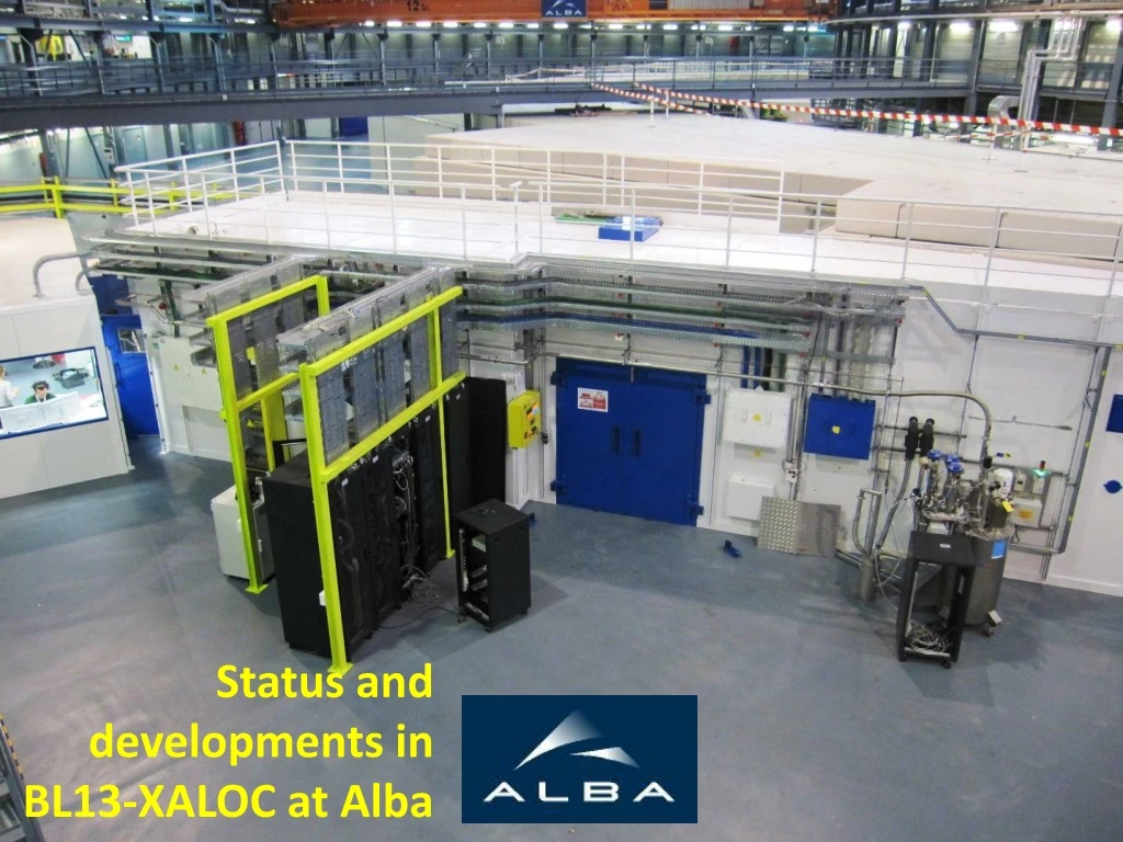
XALOC-BL13 beamline at Alba determines the 3D structure of macromolecules (proteins, DNA/RNA) using X-ray crystallography. It utilizes a diffractometer, detector, flux congelator, and microscope.

E N D
Why XALOC Robot Posiciona la mostra Difractòmetre Orienta la mostra Detector Flux congelador a -173º C Microscopi XALOC-BL13 beamline determines the 3D structure of macromolecules (proteins, ADN/ARN) using X-ray crystallography 0,1 mm Detector Cabina Experimental de Xaloc-BL13
3. Solve a X-ray diffraction pattern of protein crystal at the pixel detector Protein crystal 3D image at atomic resolution of a protein+DNA
High demand • User Operation Cycle 1 (2012+) Cycle 2 (2013) 97 BT days 53 BT days 34 Udays (29 proposals) ~50 proposals
Beamline status #5 #7 #3 #9 #11 #13 Flux (ph/s/250mA) E (keV) Beamline status : Operative (but not completed yet) Very positive comments from all (!) users • Typical beam size at sample is 5010 um FWHM (HV) as seen on a 20-um fluorescence screen • Unfocused and defocused modes were tested although not currently used. • Very reliable beam optics (alignment done daily in 10’) • Beam at sample drifts (typically downwards 1 um/hour) • Beam recovered easily after a shutdown. • Beam aligned using EH positioning tables. Optics (mirror pitches) could also be used. • Measured beam flux is >2·1012 ph/s/250mA 5010 mm (FWHM, HV) Underlying: diagnostics are very important
Beamline description Beamline concept : In-vacuum Undulator IVU21.6 + Channel-cut Si(111) monochromator + KB focusing mirrors Beamline status : Operative Mono- chromator Vertical Focusing Mirror Horizontal Focusing Mirror Diffractometer Detector Distance from source (m) 20.100 22.800 Sample changer 24.785 Dv = 3.47 Dh = 5.4 29.375 Experimental Hutch
XALOC Layout 29.375 m Detector Diffractometer 24.785 m Dh = 5.4 Photon shutter Horizontal Focusing Mirror 22.800 m Dv = 3.47 20.100 m Vertical Focusing Mirror Sample changer Monochromator Attenuator Other optical elements Fluorescence screens Diode XBPMs Slits FE trigger unit
Optics • Channel cut Si(111), 6 mm gap, by CINEL (crystals by Crystal Scientific) • 5-22 keV, with 1 mm excursion (Pt-K →Se-K: 30 mm; complete Se-MAD: 11 mm) 2nd xtal pitch (stepper motor) 2nd xtal pitch V feedback (piezo) Bremsstrahlung block I0 diode beam channel cut crystal cryogenic pipes accelerometer
Optics: mirrors • 2 mirrors, in a Kirkpatrick-Baez configuration • Manufactured by IRELEC, surfaces made by InSync • Elliptically bent using 2 motors, equipped with strain gauges • Equipped with accelerometers to monitor pitch vibrations • Fully installed Vertical Focusing Mirror Horizontal Focusing Mirror
Lysozyme data set at Rsym (inner shell)<5% High quality data: Lysozyme • Test lysozyme crystal at 100K. • Excellent overall statistics: Rsym = 3.8 %(90 deg) • Also at the lowest resolution shell (2.76A): Rsym= 2 %. • Resolution 1.26 Å. • Molecular replacement rendered excellent ED maps. lysozyme (a=b=79 Å c=37 Å; P43212) sample-det distance=200 mm l=0.9795 Å, exp time =1 s, Dw = 1° Atomic resolution data: T98D-SH3 domain of c-Src Tyr kinase • P3121 , a = b =31.5 Å, c = 106.8Å • First data set collected at 12.6 keV and at 0.93 Å resolution: • 0.5 deg oscillation, 90 deg, 1.3 sec exp time • Rsym = 3.8 %(90 deg) • Second data set collected at 15 keV and at 0.96 Å resolution: 0.5 deg oscillation, 205 deg, 2.6 sec exp time • Rsym= 3.9 % P312131.5 31.5 106.8 Crystals from A. Cámara, UA, Almería
Solve a structure using MAD at Se-K edge SAD data collection: meganuclease I-Dmol D. Mobilis • First ab initio solved structure, by Se K-edge SAD • Protein-DNA complex (PDB 2VS8) • Se-Met SAD 360˚, 2.4 Å resolution, 1 sec exp time • P21, a = 106.6, b = 70.3, c = 107.1, b = 119.9 Protein DNA Fluorescence scan widget Crystals from G. Montoya, CNIO, Madrid
Solve a structure with a unit cell par. a>400 Å Human Rhinovirus 2 • Monoclinic cell • a = 470 Å, b= 376 Å, c = 465 Å • = 90˚, = 99˚, = 90˚ • Diffraction to 4 Å. • Crystal-detector distance 750 cm • (max. distance 1350 cm) Crystals from N. Verdaguer, IBMB, Barcelona
Detectors at the Experimental Hutch Fluorescence Si-drift detector Diamond X-ray position monitors Pixel main detector PIN Diodes and YAG fluorescence screens
Detector • Pilatus 6M (Dectris) • Photon counting pixel detector • Preliminary tests on integration into BL successfully passed • Currently ending manufacturing phase • Estimated delivery in May 2011 • Data transfer and storage system to be defined ALBA turn = 893 ns 124 ns
Detector. What else? • 320 um Silicon: needed thicker • Smaller pixel size • Tiling is tolerable, but gaps should be reduced • 2.3 ms dead time: can be smaller? • Area, area • Triggers, triggers, triggers ALBA turn = 893 ns 124 ns ? ?
Other projects • Transmissive PIN diode • Collaboration with CNM • 10x10mm2 • transmissive (10 um thick) • Pinhole • Heavy Z material, 80 um thick • Hole 3-20 um diameter • High precision tools • Sharp tips <1um • Slit blades • Fluorescence dot • Mounted in a sample holder • 1 um diameter • Used to position beam and spindle
The XALOC Team Engineering Carles Colldelram Mech Engineer Alex Enrique Mech Technician Fabien Rey Alignment Marcos Quispe Cryogenics Computing Guifré Cuní Controls Engineer Oscar Matilla Electronic Engineer Julio Lidón Xavi Fariña Elec technician Experiments Jordi Juanhuix Jordi Benach Fernando Gil J.C. Martínez metrology Josep Nicolás mirrors, metrology Igors Sics diagnostics First data set April 2012 First Official Users 18 July 2012 Thank you for your attention