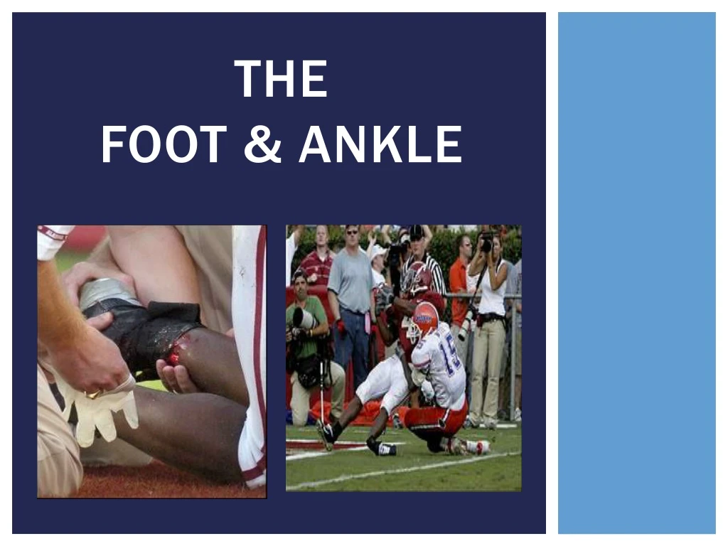
Download
1 / 160
1.6k likes | 1.64k Views
Learn the detailed anatomy of the foot and ankle, including bones, ligaments, muscles, blood vessels, and nerves. Understand important structures and functions to enhance your knowledge.

E N D
Objectives STUDENTS will be able to Identify… • The bones of the foot & ankle • The ligaments of the foot & ankle • The muscles of the foot & ankle • The blood vessels & nerves of the foot & ankle • Other structures
THE BONES • The foot contains 26 bones
The bones • The foot contains 26bones • Phalanges (14)
The bones • The foot contains 26bones • Metatarsals (5)
The bones • The foot contains 26bones • Tarsals (7)
The bones • The ankle/lower leg contains the: • Tibia • Fibula
Phalanges • There are 14 total phalanges • Great toe = 2 • All other toes = 3 • Each toe has a proximal, intermediate, & distal phalange (except Great toe)
metatarsals • Each toe has a corresponding metatarsal (MT) • Important areas: • Head of the 1st MT • Base of the 5th MT • In-between the 2-3 MT heads
Tarsal bones • There are 7 Tarsal Bones in the foot. • Cuneiforms (E, F, G) • Medial (G) • Intermediate (F) • Lateral (E) • Cuboid (C) • Navicular (D) • Talus (B) • Calcaneous (A)
Bones of the leg • Tibia and fibula extend past the talusbone • Distal end of each bone is referred to as the malleolus • Medial Malleolus = tibia • Lateral Malleolus = fibular
Bones of the leg • Tibia • Larger of the two bones • Primary weight bearing bone
Bones of the leg • Fibula • Smaller bone, extends more distally • Provides for muscle attachments • ≤ 10% weight bearing
VIDEO Foot Anatomy Ankle Anatomy
Articulations (JOINTS) • Articulation = Joint • Joint = Articulation • They can be used interchangeably • When two bones come together, they form a joint.
Subtalar joint • Articulation of the talus& calcaneus
Talocrural joint • Articulation of the tibia, fibula, talus • Also known as the Ankle Mortise.
the ligaments • Important ligaments in the foot: • Calcaneonavicular ligament • Also known as the “spring ligament”
The ligaments • Medial ankle: • Deltoid ligament
The ligaments • Lateral ankle: • Anterior talofibular(ATF) • Calcaneofibular(CF) • Posterior talofibular(PTF)
Arches of the foot • Support body weight • Absorb forces from the ground • Provide space for blood vessels, tendons, & muscles
Arches of the foot • Metatarsal Arch – across metatarsal heads • Transverse Arch – across metatarsal bases & cuneiforms • Medial Longitudinal Arch – along the medial aspect • Lateral Longitudinal Arch – along the lateral aspect
Plantar fascia • Broad, thick tissue covering the bottom of the foot • Extends from the calcaneus to the base of each metatarsal • Supports the foot against downward forces
Muscles & tendons • Intrinsic muscles of the foot: • Toe extensors • Toe flexors • Great toe & 5th toe abductors • Great toe adductor
Muscles & tendons • Extrinsic muscles of the foot/ankle: • Divided by compartments • Anterior • Lateral • Superficialposterior • Deep posterior
Muscles & tendons • Anterior compartment • Tibialis anterior • Action: • Dorsiflexion the foot
Muscles & tendons • Lateral compartment • Peroneus longus • Peroneus brevis • Action: • Eversion of the foot
Muscles & Tendons • Superficial posterior compartment • Gastrocnemius • Soleus • Action: • Plantarflexion of the foot
Muscles & tendons • Deep posterior compartment • Tibialis posterior • Action: • Plantarflexion & inversion the foot • *Muscle associated with shin splints.
Muscles & tendons • Achilles Tendon • Tough band of fibrous tissue • Connects the posterior calf muscles (Gastrocnemius and soleus) to the calcaneus
NEUROLOGICAL SUPPLY • Nerves of the foot/ankle: • Tibial nerve • Posterior leg & plantaraspect of foot • Common peroneal nerve • Anteriorleg & foot
VASCULAR SUPPLY • Blood supply • Anterior tibial artery Dorsal pedal pulse • Posterior tibial artery Posterior tibial pulse
Distal pulse Posterior Tibial Pulse Dorsal Pedal Pulse
Objectives STUDENTS will be able to Identify… • Pertinent information to gather during a foot & ankle evaluation • Important observations to make during a foot & ankle evaluation ???
The Secondary Survey • After ruling out life-threatening injuries, we begin the secondary survey • Treat for major injuries with acute on-field care • Begins with an assessment of vital signs • Musculoskeletal Assessment DOCUMENT EVERYTHING!
The Evaluation Process H.O.P.S. Technique • History • Observation • Palpation • Special tests • Range of motion
History • What happened? • Gain information about the patient and the injury • Most critical part of the evaluation! • Past medical history • History of the present condition
History • Start with generic history questions • Chief complaint • Age • Occupation / sport / position etc. • General healthcondition • Activity level • Medications
History • History of previous injuries • What happened? • Who did you see? • What did they tell you? • How long were you out? • Has it fully resolved?
History • Mechanism of injury • How did it happen? Tension = sprain; fracture; strain Torsion = sprain; fracture Compression = contusion; fracture Shear = fracture; sprain Bending = fracture
History Ask these questions regarding PAIN • Provocation – what causes it? what makes it better? • Quality – what does it feel like? neurological symptoms? • Region – where does it hurt? can you point w/one finger? • Severity – how bad does it hurt? (0-10) • Timing – when does it hurt? how long?
History • Sounds & sensations • Did you hear any sounds? • Did you hear any pops, crackles, snaps, clicks? • Did you feelanything unusual?
History • Specific to the foot & ankle • Previous history = chronic ankle instability • Mechanism of injury = ROM(Inversion, Eversion, Plantarflexion, Dorsiflexion) • Location of pain – heel, foot, toes, arches, lateral ankle, medial ankle, etc. • Determines what is injured • Changes in activity,footwear, or training surfaces
Observation • Athlete Moving? • Position of athlete? • Conscious? • Life-threatening bleeding? • If life-threating is ruled out, go to injury site.