Download
1 / 70
710 likes | 919 Views
NOTES: CH 48 Neurons, Synapses, and Signaling. A nervous system has three overlapping functions:. 1) SENSORY INPUT : signals from sensory receptors to integration centers 2) INTEGRATION : information from sensory receptors is interpreted and associated with appropriate responses

E N D
A nervous system has three overlapping functions: 1) SENSORY INPUT: signals from sensory receptors to integration centers 2) INTEGRATION: information from sensory receptors is interpreted and associated with appropriate responses 3) MOTOR OUTPUT: conduction of signals from the integration center to effector cells (muscle cells or gland cells)
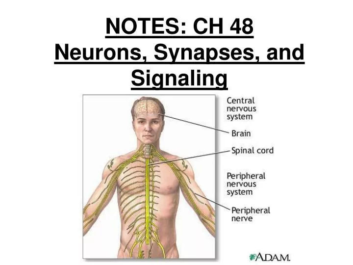
*CENTRAL NERVOUS SYSTEM (CNS) integration center brain and spinal cord
*PERIPHERAL NERVOUS SYSTEM (PNS) made up of nerves (ropelike bundles of neurons) nerves communicate motor and sensory signals to and from CNS and rest of body
Two Main Classes of Cells: 1) NEURONS: functional unit of the nervous system transmits signals from one location to another made up of: cell body, dendrites, axon many axons are enclosed by an insulating layer called the MYELIN SHEATH include: sensory neurons, interneurons, motor neurons
2) GLIAL CELLS (“GLIA”) - SUPPORTING CELLS 10 to 50 times more numerous than neurons provide structure; protect, insulate, assist neurons example: Schwann cells and oligodendrocytes form myelin sheaths in the PNS and CNS, respectively
MYELIN SHEATH: produced by Schwann cells in the peripheral nervous system; gaps between successive Schwann cells are called NODES OF RANVIER…. ***the #10 term!!!
NODES OF RANVIER! ***word #10 on my list!!! 1) Okazaki fragments 2) plasmodesmata 3) ??????? 4) ??????? 5) ??????? 6) rubisco 7) oxaloacetate 8) islets of Langerhans 9) Batesian mimicry 10) nodes of Ranvier
2) GLIA (SUPPORTING CELLS) example:astrocytes: responsible for blood-brain barrier
Astrocyte Nerve cells
ACTION POTENTIALS & NERVE IMPULSES • all cells have an electrical charge difference across their plasma membranes; that is, they are POLARIZED. this voltage is called the MEMBRANE POTENTIAL (usually –50 to –100 mV) inside of cell is negative relative to outside arises from differences in ionic concentrations inside and outside cell
**A- = group of anions including proteins, amino acids, sulfate, phosphate, etc.; large molecules that cannot cross the membrane and therefore provide a pool of neg. charge that remains in the cell
How is this potential maintained? the sodium-potassium pump uses ATP to maintain the ionic gradients across the membrane (3 Na+ out; 2 K+ in)
the “resting potential” of a nerve cell is approx. –70 mV neurons have special ion channels (GATED ION CHANNELS) that allow the cell to change its membrane potential (a.k.a. “excitable” cells)
when a stimulus reaches a neuron, it causes the opening of gated ion channels (e.g.: light photoreceptors in the eye; sound waves/vibrations hair cells in inner ear)
HYPERPOLARIZATION: membrane potential becomes more negative (K+ channel opens; increased outflow of K+) DEPOLARIZATION: membrane potential becomes less negative (Na+ channel opens; increased inflow of Na+) **If the level of depolarization reaches the THRESHOLD POTENTIAL, an ACTION POTENTIAL is triggered.
ACTION POTENTIALS (APs): the nerve impulse all-or-none event; magnitude is independent of the strength of the stimulus
5 Phases of an A.P.: 1) Resting state 2) Depolarizing phase 3) Rising phase of A.P. 4) Falling phase of AP (repolarizing phase) 5) Undershoot
Phase of A.P. State of Voltage-Gated Sodium (Na+) Channel State of Voltage-Gated Potassium (K+) channel Activation gate Inact. Gate Entire channel 1) Resting closed open closed closed 2 & 3) Depolari-zation open open open closed 4) Repolar-ization open closed closed open 5) Undershoot closed closed closed open
**during the undershoot, both Na+ channel gates are closed; if a second depolarizing stimulus arrives during this time, the neuron will NOT respond (REFRACTORY PERIOD) strong stimuli result in greater frequency of action potentials than weaker stimuli
How do action potentials “travel” along an axon? the strong depolarization of one action potential assures that the neighboring region of the neuron will be depolarized above threshold, triggering a new action potential, and so on…
SYNAPSE: junction between a neuron and another cell; found between: -2 neurons -sensory receptor & sensory neuron -motor neuron & muscle cell -neuron & gland cell