Download
1 / 28
280 likes | 328 Views
Understand stages of renal failure, workup approaches, major causes, history and exams, lab investigations, subsequent tests, imaging, biopsy, management plans, and hypertension control. Learn how to identify reversible causes, slow disease progression, treat complications, and manage renal replacement therapy. Explore the significance of hypertension control with AII inhibition and ACEI/ARBS.

E N D
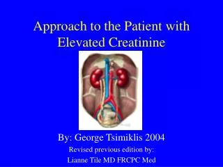
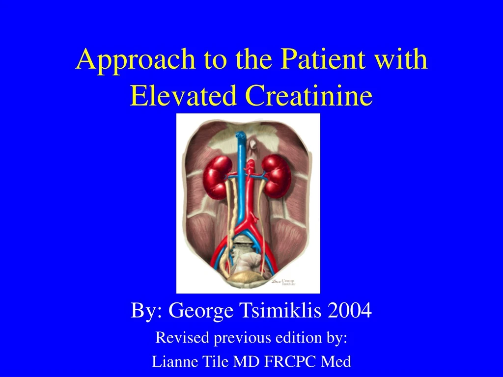
Approach to the Patient with Elevated Creatinine By: George Tsimiklis 2004 Revised previous edition by: Lianne Tile MD FRCPC Med
Objectives • To identify appropriate strategies for investigation of the patient with increased creatinine • To discuss interventions that may alter the course of disease • To discuss indications for referral to a nephrologist
Creatinine is an estimate of GFR Cockcroft-Gault: (140-age) x wt x 100 = GFR (cc/min) 72 x serum Cr GFR (females) = GFR (males) x 0.85
CASES: What is Considered an ELEVATED Creatinine? 55 yo 70 kg male with Cr of 220: 75 yo 45kg female with Cr of 220: 75 yo 45kg female with Cr of 85: 75 yo 45kg female with Cr of 45: GFR =37 moderate GFR = 16 severe GFR =40 moderate GFR =76 mild
Workup of the patient with high Cr • Approach • 1. Identify chronicity (Acute vs chronic) • 2. Identify the cause, especially reversible causes • 3. Identify Indications for Referral to a Nephrologist • 4. Initiate a cause specific management plan in a multidisciplinary team.
ACUTE Fever/ hematuria Hypovolemia Hydroureter Sepsis New hypertension Recent nephrotoxins No hypocalcemia No hyperphosphatemia No anemia CHRONIC previous confirmed nephropathy Already diminished CrCl Atrophic kidneys (<10cm on U/S) Normochromic normocytic anemia Hypocalcemia Hyperphosphatemia Acute vs Chronic Renal Failure
Major Causes of Renal Failure • Prerenal: decreased glomerular perfusion • volume depletion: diuretics, poor intake • decreased effective circulating volume: forward heart failure, cirrhosis, sepsis • Renal • Vascular disease: acute (vasculitis, thromboembolic, HUS/TTP, malignant hypertension) vs. chronic (renal artery stenosis, HTN/nephrosclerosis) • Glomerular disease: nephritic vs. nephrotic • Tubular/interstitial disease: acute (ATN, AIN, myeloma) vs. chronic (PCKD, pyelo, autoimmune, analgesic abuse) • Post-Renal/ Obstructive Nephropathy • Malignancy, prostate hyperplasia/ cancer
History and Physical Exam • signs or symptoms of • underlying disorder: i.e. volume status, flank pain, obstruction, diabetes, hypertension, vasculitis • altered kidney function: urine output, urine discoloration, edema • renal failure: anorexia, vomiting, altered mental status, HTN • medications: NSAID, ACEI, analgesics, aminoglycosides, contrast, Chinese herbs
Initial Laboratory Investigations • Urinalysis: hematuria, pyuria, proteinuria • Urinary Sediment: casts, GN • Urine Volume: oliguric, obstruction • 24-hour Urine protein and CrCl • Urea: • Electrolytes • CBC: thrombocytopenia: TTP, HUS; Anemia: cause or effect of renal disease; Leukocytosis: ?pyelo, infection • Glucose: DM? • Bicarbonate: metabolic acidosis • Calcium and Phosphate • Protein and albumin • Serum protein electrophoresis
Subsequent Laboratory Investigations Blood Tests • Immunity Studies: ANA, anti-DNA, RF, ANCA, C3, C4 • Inflammatory Studies: ESR, CRP • Serology: Hep B, HIV, Bence-Jones • Urinary Eosinophilia: AIN, embolic disease • CK • Liver enzymes, hepatitis: secondary GN
Imaging • Ultrasound • rule out obstruction, stones, mass • small kidneys suggest chronic process • normal sized kidneys do not exclude chronic disease (amyloid, DM,myleoma, PCKD). • Doppler may be used to assess blood flow of arteries (RAS) or veins (thrombosis) • CT Scan - useful for stones and masses • If suspected renal artery stenosis: MR angiography (no nephrotoxic dye), renal angiogram
Renal Biopsy • Should be considered: • if noninvasive tests have failed to establish a diagnosis in a patient with: • nephrotic syndrome (except in DM) • Certain cases of non-nephrotic proteinuria if associated with renal dysfunction • Lupus nephritis (for dx and staging) • acute nephritic syndrome • unexplained acute/ subacute renal failure • to differentiate GN from vasculitis • to direct and evaluate effectiveness of therapy
Management of Renal Disease • Treatment of Reversible Causes • Preventing or Slowing Progression • Hypertension Control (<130/80) • Control Proteinuria (<500-1000mg/day or 60% from baseline values) • Treating and Preventing the Complications • Identifying Individuals Requiring Renal Replacement Therapy
Hypertension • Controlling BP slows progression of disease • target <125/80, lower if normotensive at baseline or with more proteinuria • Slowing of disease is related to decreased systemic BP, decreased glomerular hypertension, and decreased urine protein excretion • Microalbuminuria is a major cardiovascular risk factor!
AII Inhibition • ACEI/ ARBS are more effective than other medications in slowing progression: • Also benefit normotensive diabetics • Secondary actions include decrease glomerular remodelling • preferential antiproteinuric effect • decreases AII effects • slows the progression of renal disease in type 1 and 2 DM independent of the effect on BP • in nondiabetics, most convincing effect is in patients with greater proteinuria
Other Antihypertensives • ARB: • benefit similar to ACEI seen in DM-2 • may be additive benefit with ACEI • Verapamil, Diltiazem or beta-blocker • antiproteinuric effect in diabetics • Other medications to achieve BP goals
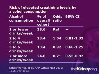
Diet: Protein Restriction • Dietary protein restriction (0.4-0.6 g/kg/day) reduced the risk for renal failure or death in nondiabetic renal disease and slowed nephropathy in type I DM • beneficial effects were unrelated to change in blood pressure or glycemic control. • NNT 5 to 50 • realistic to restrict to 0.8-1g/kg/day Ann Intern Med. 1996 April 1;124: 627-32.
Dyslipidemia • Abnormal lipid metabolism is common in RF • Primary problem is hypertriglyceridemia • LDL, HDL, chol may also be deranged • CRF is considered a high cardiovascular risk and therefore target lipid levels are as those with CAD • Some suggest that statins may benefit the kidney independent of lipid lowering effects
Erythropoietin • Indicated for Hgb <110 • Chk iron stores, folate, and B12 • In patients with ferritin <100 and sat <20%, must supplement prior to commencement of therapy and continue supplement while on EPO • Follow response to EPO and ensure adequate residual stores
Case • Your patient returns for follow-up after 6 months. He has no new symptoms. You repeat his serum creatinine. It is now 185 (from 155). • At what creatinine level would you refer him to a nephrologist? a) Now (185) b) 250 C) 310
Referral to Nephrologist • Late referral (< 12 months pre dialysis) is common • Survey of Ontario Family MDs: • 84% would not refer with creat 120-150 (>50% loss of GFR) • 28% would not refer with creat 150-300 • almost all would refer with creat>300 • Consequences of referral shortly before dialysis: • more complications • longer hospitalization to initiate dialysis • more difficulty with initiation of dialysis • worse survival! • Better outcomes with early multidisciplinary care CMAJ 1999: 161:413-17
Canadian Guidelines • Renal replacement therapy is NOT rationed (i.e. everyone should be considered) • Reversible causes should be sought at diagnosis • At least 1 year is required to prepare for dialysis • Refer, at the latest, at Cr clearance of 30 ml/min, or Cr of 300 • But…there are probably not enough nephrologists/ clinics to meet this demand • Adequate communication with the Nephrologist will allow proper stratification of patients CMAJ 1999: 161:413-17
For AIMGP Clinic • It is reasonable to follow stable renal failure patients, and work up and manage appropriately • Refer to nephrology when: • Cr >300 or Cr clearance <30 ml/min • Renal biopsy indicated • Indicators of aggressive disease are present: • Rapid decline in creatinine • homeostatic derangement i.e. acidosis, volume overload, high K • high protein excretion • Difficult to control BP • low HDL • black race
Conclusions • When evaluating a patient with increased creatinine, internists should: • Identify and treat reversible causes of renal failure • Initiate management to slow the decline in renal function • Manage coexisting conditions • Have clear indications for when to refer to nephrology subspecialists
References • Elevated Serum creatinine: recommendations for management and referral. CMAJ 1999: 161:413-17 • Approach to managing elevated creatinine. Can Fam Physician. 2004;50:735-740. • UptoDate