Download
1 / 1
10 likes | 63 Views
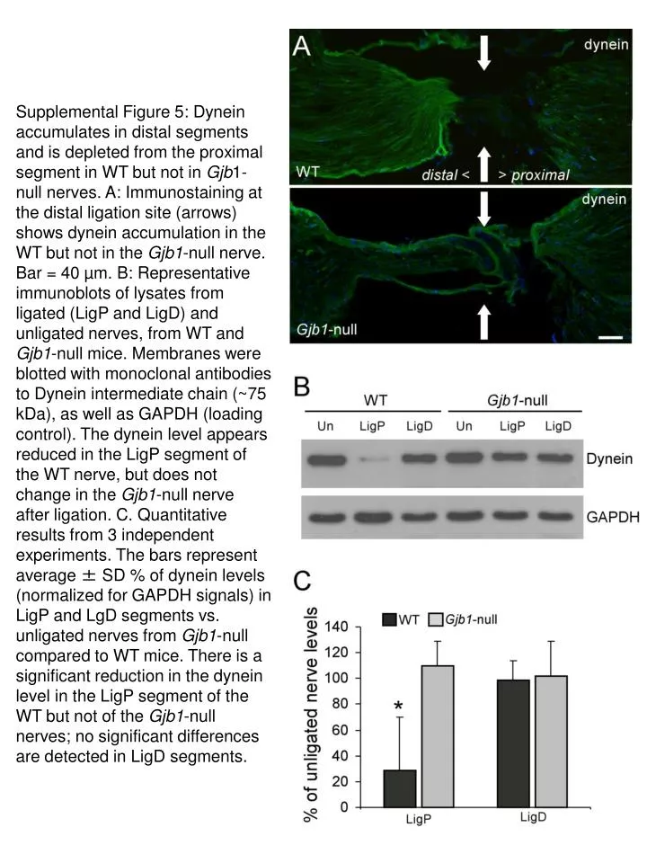
Supplemental Figure 5 shows dynein distribution in WT and Gjb1-null nerves. Dynein accumulates distally in WT but not Gjb1-null nerves. Immunoblots reveal reduced dynein levels in WT LigP segments but not in Gjb1-null nerves. Quantitative data confirms significant dynein reduction in WT LigP segment.

E N D
Supplemental Figure 5: Dynein accumulates in distal segments and is depleted from the proximal segment in WT but not in Gjb1-null nerves. A: Immunostaining at the distal ligation site (arrows) shows dynein accumulation in the WT but not in the Gjb1-null nerve. Bar = 40 µm. B: Representative immunoblots of lysates from ligated (LigP and LigD) and unligated nerves, from WT and Gjb1-null mice. Membranes were blotted with monoclonal antibodies to Dynein intermediate chain (~75 kDa), as well as GAPDH (loading control). The dynein level appears reduced in the LigP segment of the WT nerve, but does not change in the Gjb1-null nerve after ligation. C. Quantitative results from 3 independent experiments. The bars represent average ± SD % of dynein levels (normalized for GAPDH signals) in LigP and LgD segments vs. unligated nerves from Gjb1-null compared to WT mice. There is a significant reduction in the dynein level in the LigP segment of the WT but not of the Gjb1-null nerves; no significant differences are detected in LigD segments.