Download
1 / 56
570 likes | 628 Views
Explore the intricate world of cellular components, organelles, and fractionation methods. Learn about the energy-related organelles like mitochondria and chloroplasts, as well as techniques such as differential centrifugation. Gain insights into cell function and structure correlation.

E N D
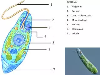
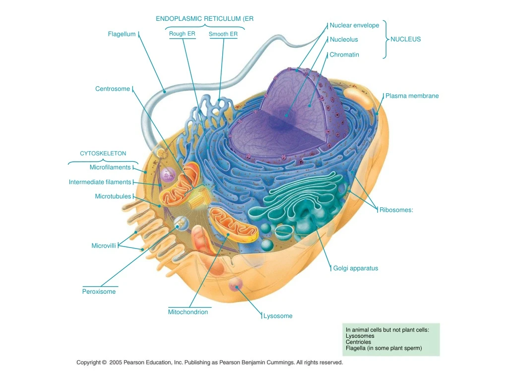
ENDOPLASMIC RETICULUM (ER Nuclear envelope Flagellum Rough ER Smooth ER NUCLEUS Nucleolus Chromatin Centrosome Plasma membrane CYTOSKELETON Microfilaments Intermediate filaments Microtubules Ribosomes: Microvilli Golgi apparatus Peroxisome Mitochondrion Lysosome In animal cells but not plant cells: Lysosomes Centrioles Flagella (in some plant sperm)
Centrifugation & Quantification • Cellular Structure • Centrifugation • Beer-Lambert Equation • Absorption Efficiency
Energy-Related Organelles:Mitochondria • Bounded by double membrane • Cristae – Infoldings of inner membrane that encloses matrix • Matrix – Inner semifluid containing respiratory enzymes • Involved in cellular respiration • Produce most of ATP utilized by the cell
Energy-Related Organelles:Chloroplast • Bounded by double membrane • Inner membrane infolded • Forms disc-like thylakoids, which are stacked to form grana • Suspended in semi-fluid stroma • Green due to chlorophyll • Green photosynthetic pigment • Found ONLY in inner membranes of chloroplast
Captures light energy to drive cellular machinery • Photosynthesis • Synthesizes carbohydrates from CO2 & H2O • Makes own food using CO2 as only carbon source • Energy-poor compounds converted to enery rich compounds • Chloroplast structure includes: • Thylakoids, membranous sacs, stacked to form a granum • Stroma, the internal fluid
Purification of Cell Parts • Understanding the roles of each cell component depends on methods to break (lyse) cells and separate cell components for analysis • Cell lysis is accomplished by various techniques: blender, sonication, tissue homogenizer, hypotonic solution • Separation of cell components generally involves centrifugation
Cell Fractionation • Cell fractionation takes cells apart and separates the major organelles from one another • Ultracentrifuges fractionate cells into their component parts • Cell fractionation enables scientists to determine the functions of organelles • Biochemistry and cytology help correlate cell function with structure
Centrifugation A centrifuge is used to separate particles or even macromolecules: - Cells - Sub-cellular components - Proteins - Nucleic acids Basis of separation: - Size - Shape - Density Methodology: - Utilizes density difference between the particles/macromolecules and the medium in which these are dispersed - Dispersed systems are subjected to artificially induced gravitational fields
Homogenization Tissue cells Homogenate Differential centrifugation
axis of rotation Swinging-bucket g At rest Spinning g Fixed-angle Centrifuge Rotors
axis of rotation rmax rav rmin a rmin rmin rav rav rmax rmax b c Sedimentation path length Geometry of Rotors
v = velocity of sedimentation d = diameter of particle l = density of liquid p = density of particle = viscosity of liquid g = centrifugal force Stokes Equation
Types of Centrifugation Differential Centrifugation: Rate-Zonal Centrifugation Isopycnic Centrifugation: Density Equilibrium Centrifugation
Differential Centrifugation • This is the most common method of fractionating cells • Fractionation is the separation of the different organelles within the cell
Differential Centrifugation • Density of liquid is uniform • Density of liquid << Density of particles • Viscosity of the liquid is low • Consequence: Rate of particle sedimentation depends mainly on its size and the applied G-force.
Size of Major Cell Organelles • Nucleus 4-12 m • Plasma membrane sheets 3-20 m • Golgi tubules 1-2 m • Mitochondria 0.4-2.5 m • Lysosomes/peroxisomes 0.4-0.8 m • Microsomal vesicles 0.05-0.3m
Decant supernatant 1000g/10 min 3000g/10 min etc. Differential Centrifugation of a Tissue Homogenate
1,000 g (1,000 times the force of gravity) 10 min Supernatant poured into next tube 20,000 g 20 min 80,000 g 60 min Pellet rich in nuclei and cellular debris 150,000 g 3 hr Pellet rich in mitochondria (and chloro-plasts if cells are from a plant) Pellet rich in “microsomes” (pieces of plasma membranes and cells’ internal membranes) Pellet rich in ribosomes
Differential Centrifugation Poor resolution and recovery because of: • Particle size heterogeneity • Particles starting out at rmin have furthest to travel but initially experience lowest RCF • Smaller particles close to rmax have only a short distance to travel and experience the highest RCF
Swinging-bucket rotor: Long sedimentation path length gmax >>> gmin Fixed-angle rotor: Shorter sedimentation path length, gmax > gmin Differential Centrifugation
Density Barrier Discontinuous Continuous Density Gradient Centrifugation
Least dense Most dense How Does a Gradient Separate Different Particles?
Stokes Equation When p > l : v is +ve When p = l : v is 0 When p < l : v is –ve
1 2 3 4 5 Buoyant density banding Equilibrium density bandingIsopycnic banding
1 2 3 3 Formats for Separation of Particles According to Density When density of particle < density of liquid V is -ve
Density Barrier Discontinuous Continuous I II Resolution of Density Gradients
Isopycnic Centrifugation The density gradient is formed during the centrifugation. The sample is dissolved in solution of dense salt (e.g., CsCl, Cs2(SO4)) The macromolecules of the biological sample seek an area in the tube where the density is equal to their respective densities
Separation of Particles According to Size p >> l : v is +ve for all particles throughout the gradient
SAFETY during Centrifugation A rotor spinning at high speed is DANGEROUS Always balance the tubes, to avoid damaging the centrifuge or yourself. Never open a centrifuge that is spinning Centrifuges generate aerosols
Organelle Separation Procedure • Cut tissue in an ice-cold isotonic buffer. It is cold to stop enzyme reactions, isotonic to stop osmosis and a buffer to stop pH changes. • Grind tissue in a blender to break open cells. • Filter to remove insoluble tissue
Quantitation of Concentration • UV/vis Absorption • Limitation of Beer-Lambert Equation • Correct Choice of Wavelength • Spec 20
cuvette slit source detector Beer’s Law • A = -logT = log(P0/P) = ecL • T = Psolution/Psolvent = P/P0 • Works for monochromatic light • Compound x has a unique e at different wavelengths
Color of the sample • Remember: • Solution absorbs red appears blue-green • Solution absorbs blue-green appears red
Types of Electronic Transition • Three types • Involving p, s, and n electrons • Involving d and f orbital electrons • Charge transfer electrons
Beer’s Law Analysis • Choice of wavelength • Typically choose wavelength of maximum absorbance • May deviate from this to avoid an interference
Cuvette • The cuvettes for use in the Spec 20 resemble small test tubes. • Each cuvette is marked so that it can be positioned properly in the sample holder. • The mark is at the top of the cuvette and must be positioned toward the front of the spectrophotometer when taking measurements. • Handle these tubes with extreme care to keep both the inside and outside surfaces clean and free of scratches. • Cleaning the Cuvette: • NEVER use a brush to clean the inside of the cuvette. • Rinse the tube with distilled water a few times • Add about 1 mL of the solution to be measured. Tilt and turn the cuvette so that the solution has contact with all the surfaces. Discard this solution and repeat this rinse once more. • Fill the cuvette about 3/4 full of the solution you wish to test. • Wipe the outside of the cuvette with a lint-free, soft tissue (a Chemwipe) to remove any moisture or fingerprints from the outside surface.