Download
1 / 46
490 likes | 819 Views
SURGICAL TREATMENT DIABETIC FOOT INFECTIONS. Lidia Ionescu, Cozmin Radulescu, Daniel Guta, Irina Trifescu cl.III chirurgie, UMF Iasi. Epidemiology. The prevelence of DM –rising dramatically worldwide Largely related to increasing rates of obesity WHO- 5% of the world’s pop. DM- 2025

E N D
SURGICAL TREATMENTDIABETIC FOOT INFECTIONS Lidia Ionescu, Cozmin Radulescu, Daniel Guta, Irina Trifescu cl.III chirurgie, UMF Iasi
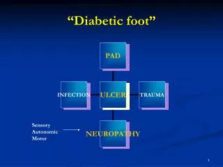
Epidemiology The prevelence of DM –rising dramatically worldwide Largely related to increasing rates of obesity WHO- 5% of the world’s pop. DM- 2025 The rising prevalence+increased longevity of pop.- rise in DM-associated complications. One of the most feared and frequent- amputation The sequence: ulceration of an insensate, diformed foot- wound infection
DM-associated foot infections Considerable morbidity Occasionally mortality- gas gangrene-septic shock Repeated and prolonged hospital admissions Costly treatment
Prospective study, USA, DM-care setting • 9% foot infection-2 years of follow-up, despite: • Educational sessions • Therapeutic shoes • Follow-up in a foot clinic • Ready access to podiatric care
DM foot infectionlower extremity amputation • DM-underlying cause of 60% amputations in developed countries • Norway study-the rate of amputation 32 times higher than among non-DM pts. • Postop. mortality: 10%-15%: • advanced age, • coronary and peripheral vascular disease, • renal failure
Risk Factors for Foot Ulceration and Infection Risk factor Mechanism of injury or impairment Peripheral motor neuropathy Abnormal foot anatomy and biomechanics, with clawing of toes, high arch, and subluxed metatarsophalangeal joints, leading to excess pressure, callus formation, and ulcers Peripheral sensory neuropathy Lack of protective sensation, leading to unattended minor injuries caused by excess of pressure or mechanical or thermal injury Peripheral autonomic neuropathy Deficient sweating leading to dry, cracking skin Neuro-osteoarthropathic deformities Abnormal anatomy and biomechanics, leading to excess pressure, especially in the midplantar area Vascular insufficiency Impaired tissue viability, wound healing, and delivery of neutrophils Metabolic derangements Impaired immunologic (especially neutrophil) function and wound healing, and excess collagen cross-linking Patient disabilities Reduced vision, limited mobility, and previous amputation Maladaptive patient behaviors Inadequate adherence to precautionary measures and foot inspection and hygiene procedures, poor compliance with medical care, inappropriate activities, excessive weight-bearing, and poor footwear Health care system failures Inadequate patient education and monitoring of glycemic control and foot care
Microbiology • Obtaining proper specimens, avoid: • Missing true pathogens • Isolating contaminating organisms
Recommandations for the methods of collecting and processing wound specimens for culture • Debride and cleanse the wound before taking a specimen for culture • Obtain tissue specimens from ulcers by curettage • Aspirate purulent secretions • Biopsy deep tissue or bone infections • Avoid sending wound swab for culture • Obtain blood cultures if patient is seriously ill • Label and send specimens promptly in sterile containers or transport media for aerobic and anaerobic cultures • Request Gram-stained smear of specimen
Management • Foot infections- potentially catastrophic outcome • Management must be: • timely, • rational, • well coordinated.
Key points Initial assessment- severity and extent of the infection Ischaemia of the affected limb confers a poor prognosis, the vascular status must be delineated early in the assessment of the inf. It can be difficult to assess the extent of an inf. without carrying out appropriate debridement, with the goal of removing all necrotic tissue Drain abscesses or remove necrotic soft tissue Antibiotics are necessary but rarely sufficient for treating diabetic foot inf., they are best seen as adjunctive not primary, therapy in treating these infections Coordinated, multidisciplinary approach: diabetologist, infection specialist, vascular surgeon, plastic surgeon,podiatrist.
Grading the severity of infection • When a pt. presents with a foot wound, the clinician should determine: • whether or not it is infected • if infected, how severely • If hospital admission is needed • If urgent surgery is required • If parenteral and broad-spectrum antibiotics are indicated
Clinical classificationIDSA • Mild infection: present pus or > 2 signs of inflammation but: • extent of cellulitis<2 cm. around the wound • limited to skin and superf. sc. tissues, • no other local complications. • no systemic illness
Clinical classification IDSA • Moderate infection: inf. in systemically well and metabolically stable pt. who has: • cellulitis extending>2cm., • lymphangitis, • spread beneath the superf. fascia, • deep tissue abscess, • gangrene, • involvement of muscle, tendons, joint, bone
Clinical classification IDSA • Severe infection: infection in a pt. with systemic toxicity or metabolic instability: • fever, chills, • tachycardia, • hypotension, • confusion, • leukocytosis, • acidosis, • severe hyperglycaemia, • azotaemia
Foot infections Foot ischeamia may increase the severity of any infection The rates of hospitalization and amputation increase with infection severity
Therapy • The goals of therapy for patients with diabetic foot infection are : • the eradication of clinical evidence of infection • the avoidance of soft tissue loss and amputations. • Good clinical response can be expected in 80%- 90% of mild to moderate infections and in 60%- 80% of severe infections or in cases of osteomyelitis. • Relapses occur in 20%- 30% of patients.
Role of debridement in assessment and treatment • Removal of non-viable tissue and surrounding callus to eliminate a sourse of bacterial colonization in the underlying tissue • Properly done, allows full assessment of the extent and depth of ulceration and tissue necrosis • Adequate debridement must precede the application of topical wound-healing agents, and dressings.
Role of debridement in assessment and treatment Debridement on a regular basis is thought to improve the rate of wound healing Failure of the treating clinician to adequately debride a wound: - lack of training, - lack of knowledge , - lack of time, is a frequent cause of healing failure.
Role of surgery in treatment of infection • The need for surgery must be carefully considered early in the evaluation process • Mild infections- surgery unlikely • Severe infections • excision of necrosis until healthy, bleeding tissues are encountered • Pus must be drained • Joint resection/partial amputation of the foot may be needed: osteomyelitis, septic arthritis, gangrene.
Emergency surgery Aim- to stop the progression of infection Amputations to the level of viable soft tissue and bone All post-surgical pts. require careful podiatric follow-up and attention to any orthotic or prosthetic needs.
Dressings • Many dressings on the market: • Hydrogels- dry to minor draining wounds • Hydrocolloids- moderate draining wounds • Polyurethane foams- superabsorbent • Calcium alginates- heavy exudative wounds • Collagen dressings- heavily draining wounds • Antimicrobial dressings (silver, iodine) • Skin replacements • Vacuum-assisted closure dressings- negative pressure wound therapy
Dressings • To facilitate healing: • Should prevent desiccation • Absorb excess fluid • Protect the wound from contamination and trauma
Dressings • The type of the dressings selected depends upon: • Wound’s size • Depth • Location • Surface characteristics
Case report PM, 69 years old, Vetrisoaia-Husi, DM-type I-for 34 years Emergent hospital admission- april 2004 Dg.- severe sepsis, plantar necrotizing fasciitis Referred from another hospital, where he refused amputation. 1 week history of acute inflammatory signs around an old foot ulcer Surgical treatment- limited plantar incisions- extended infection+systemic complications.
Case report • On admission: fever,chills, confused, pale, poor urinary output but cardio-vascular stable • Local examination: • 2 plantar incissions and retromaleolar, foul smelling secretions, necrotic subcut. tissues • Above knee cellulitis • Edema of the thigh and external genitalia organs • Inguinal lymphadenopathy • Peripheral pulses, present but weak
Case report • Severe septis: • WBC=33200/mmc • Sec. anemia, Hb=8,2g/dl., Ht=25% • Hyperpgycemia-256mg/dl • Renal failure-urinary output<50ml./h. • Urea=76mg/dl, creatinine=1,13mg/dl • Hyponatremia=129mEq/l
Case reportTreatment • Correction of deficits • Antibiotherapy iv- quinolone+flagyl • G-stained smear of specimen=+/-G cocci. • Emergent surgical treatment under iv GA: • Enlargement of previous incisions • Debridements till muscular layer Cultures of necrotic tissue- enteroccocus Persistent fever and high WBC- infectionist- imipeneme 4g/day, 10 days
Case report Repeated surgical debridements After 3 weeks- clean wounds, granulating tissue But, large soft-tissue loss at the sole After 1 month- repeated skin grafting After 1 month-plastic surgeon- cross-leg to cover the calcanean region defect After 3 weeks-separation of the cross-leg
Case report Hospital stay- 4 months and 3 weeks Large amounts of dressings Time-consumer treatment Costly treatment BUT, the leg was saved and the patient can walk and work in agriculture.