Download
1 / 16
190 likes | 537 Views
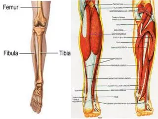
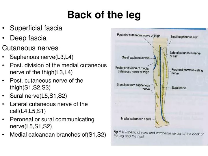
Back of the leg. Superficial fascia Deep fascia Cutaneous nerves Saphenous nerve(L3,L4) Post. division of the medial cutaneous nerve of the thigh(L3,L4) Post. cutaneous nerve of the thigh(S1,S2,S3) Sural nerve(L5,S1,S2) Lateral cutaneous nerve of the calf(L4,L5,S1)

E N D
Back of the leg • Superficial fascia • Deep fascia Cutaneous nerves • Saphenous nerve(L3,L4) • Post. division of the medial cutaneous nerve of the thigh(L3,L4) • Post. cutaneous nerve of the thigh(S1,S2,S3) • Sural nerve(L5,S1,S2) • Lateral cutaneous nerve of the calf(L4,L5,S1) • Peroneal or sural communicating nerve(L5,S1,S2) • Medial calcanean branches of(S1,S2)
Back of the leg Flexor retinaculum Anteriorly- posterior border and tip of the medial melleolus Posterioly and laterally- medial tubercle of the calcaneum
Back of the leg Structures passing deep to the flexor retinaculum:- • Tibialis posterior • Flexor digitorum longus • Post. Tibial artery • Tibial nerve • Flexor hallucis longus
Back of the leg Superficial muscles- Gastrocnemius: Origin-medial head arises from medial condyle of the femur, behind the adductor tubercle, popliteal surface of the femur and capsule of the knee jt. lateral head- lateral condyle of the femur, lateral supracondylar line and capsule of the knee jt. Insertion- fuses with muscles of the soleous to form tendocalcaneus or Achilles-middle 1/3rd of the post. Surface of the calcaneum Nerve supply-tibial nerve
Back of the leg Soleus- Origin-Fibula: back of the head and upper 1/4th of the post. Surface of the shaft tibial: soleal line and middle 1/3rd of the medial border of the shaft Insertion- Same as that of the gastrocnemius Nerve supply- Tibial nerve
Back of the leg Plantaris- Origin: Lower part of the lateral supracondylar line of the femur and oblique popliteal ligament Insertion-post surface of the calcaneum, medial to the tendocalcaneus. Nerve supply- Tibial nerve
Back of the leg Deep muscles: Popliteus- Origin: ant part of the popliteal groove on the lateral surface of the lateral condyle of the femur, arcuate popliteal lig and outer margin of the lateral meniscus of the knee jt. Insertion: post surface of the tibia Nerve supply-tibial nerve
Back of the leg Flexor digitorum longus- Origin: upper 3/4th of the medial part of the post surface of the tibia below the soleal line Insertion: 4 tendons into lateral 4 toes. Nerve supply-tibial nerve
Back of the leg Flexor hallucis longus- Origin: lower 3/4th of the post surface of the fibula and interosseous membrane Insertion: base of the distal phalanx of the great toe. Nerve supply- tibial nerve
Back of the leg Tibialis posterior- Origin: upper 2/3rd of the lateral part of the post surface of the tibia below the soleal line, post surface of the fibula infront of the medial crest and post surface of the interosseous membrane Insertion-tuberosity on the navicular bone and also it gives slips to all the tarsal bones except talus,and 2nd and 3rd metatarsal bone Nerve supply- Tibial nerve
Back of the leg Post tibial artery Course and relations Branches- • Peroneal artery • muscular branch • Nutrient artery • Circumflex fibular branch • Communicating branch • Malleolar branch • Calcaneal branches • Terminal branches
Back of the leg Peroneal artery Course and relation Branches- • Muscular branches • Nutrient artery • Anastomotic branches