Download
1 / 92
940 likes | 1.25k Views
Learn the primary and secondary indications of cardiac ultrasound, including assessing cardiac wall motion and detecting pericardial effusion. Explore main clinical questions, probe selection, and various cardiac views for diagnosis. This guide covers practical tips and views for a comprehensive cardiac evaluation in emergency settings.

E N D
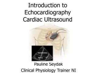
Cardiac Ultrasound in Emergency Medicine Anthony J. Weekes MD, RDMS Sarah A. Stahmer MD For the SAEM US Interest Group
Primary Indications • Thoraco-abdominal trauma • Pulseless Electrical Activity • Unexplained hypotension • Suspicion of pericardial effusion/tamponade
Secondary Indications • Acute Cardiac Ischemia • Pericardiocentesis • External pacer capture • Transvenous pacer placement
Main Clinical Questions • What is the overall cardiac wall motion? • Is there a pericardial effusion?
Cardiac probe selection • Small round footprint for scan between ribs • 2.5 MHz: above average sized patient • 3.5 MHz: average sized patient • 5.0 MHz: below average sized patient or child
Main cardiac views • Parasternal • Subcostal • Apical
Wall Motion • Normal • Hyperkinetic • Akinetic • Dyskinetic: may fail to contract, bulges outward at systole • Hypokinetic
Orientation • Subcostal or subxiphoid view • Best all around imaging window • Good for identification of: • Circumferential pericardial effusion • Overall wall motion • Easy to obtain – liver is the acoustic window\
Subcostal View • Most practical in trauma setting • Away from airway and neck/chest procedures
Subcostal View • Liver as acoustic window • Alternative to apical 4 chamber view
Subcostal View • Angle probe right to see IVC • Response of IVC to sniff indicates central venous pressure • No collapse • Tamponade • CHF • PE • Pneumothorax
Parasternal Views • Next best imaging window • Good for imaging LV • Comparing chamber sizes • Localized effusions • Differentiating pericardial from pleural effusions
Parasternal Long Axis • Near sternum • 3rd or 4th left intercostal space • Marker pointed to patient’s right shoulder (or left hip if screen is not reversed for cardiac imaging) • Rotate enough to elongate cardiac chambers
Parasternal Short Axis • Obtained by 90° clockwise rotation of the probe towards the left shoulder (or right hip) • Sweep the beam from the base of the heart to the apex for different cross sectional views
Apical View • Difficult view to obtain • Allows comparison of ventricular chamber size • Good window to assess septal/wall motion abnormalities
Apical Views • Patient in left lateral decubitus position • Probe placed at PMI • Probe marker at 6 o’clock (or right shoulder) • 4 chamber view
Apical 4 chamber view • Marker pointed to the floor • Similar to parasternal view but apex well visualized • Angle beam superiorly for 5 chamber view
Apical 2 chamber view • Patient in left lateral decubitus position • Probe placed at PMI • Probe marker at 3 o’clock • 2 chamber view
Apical 2 chamber view • Good look at inferior and anterior walls
Apical 2 chamber view • From apical 4, rotate probe 90° counterclockwise • Good view for long view of left sided chambers and mitral valve
Abnormal findings Pericardial Effusion
Case Presentation • 45 year old male presents with SOB and dizziness for 2 days. He has a long smoking history, and has complained of a non-productive cough for “weeks” • Initial VS are BP 88/palp, HR 140 • PE: Neck veins are distended • Chest: Clear, muffled heart sounds • Bedside sonography was performed
Echo free space around the heart • Pericardial effusion • Pleural effusion • Epicardial fat (posterior and/or anterior) • Less common causes: • Aortic aneurysm • Pericardial cyst • Dilated pulmonary artery
Size of the Pericardial Effusion • Not Precise • Small: confined to posterior space, < 0.5cm • Moderate: anterior and posterior, 0.5-2cm (diastole) • Large: > 2cm
Clinical features of Pericardial effusion • Pericardial fluid accumulation may be clinically silent • Symptoms are due to: • mechanical compression of adjacent structures • Increased intrapericardial pressure
Pericardial Effusion:Asymptomatic • Up to 40% of pregnant women • Chronic hemodialysis patients • one study showed 11% incidence of pericardial effusion • AIDS • CHF • Hypoproteinemic states
Symptoms of Pericardial Effusion • Chest discomfort (most common) • Large effusions: • Dyspnea • Cough • Fatigue • Hiccups • Hoarseness • Nausea and abdominal fullness
Cardiac Tamponade • Increased intracardiac pressures • Limitation of ventricular diastolic filling • Reduction of stroke volume and cardiac output
Abnormal findings • Is the cause of hypotension cardiac in etiology? • Is it due to a pericardial effusion? • Is is due to pump failure?
Unexplained Hypotension • Cardiogenic shock • Poor LV contractility • Hypovolemia • Hyperdynamic ventricules • Right ventricular infarct/large pulmonary embolism • Marked RV dilitation/hypokinesis • Tamponade • RV diastolic collapse
Cardiogenic shock • Dilated left ventricle • Hypocontractile walls
Hypovolemia • Small chamber filling size • Aggressive wall motion • Flat IVC or exaggerated collapse with deep inspiration
Massive PE or RV infarct • Dilated Right ventricle • RV hypokinesis • Normal Left ventricle function • Stiff IVC
Case presentation ? overdose • 27 yo f brought in with “passing out” after night of heavy drinking. • Complaining of inability to breathe! • PE: Obese f BP 88/60 HR 123 Ox 78% • Chest: clear • Ext: No edema • Bedside sonography was performed
Chest pain then code • 55 yo male suffered witnessed Vfib arrest in the ED • ALS protocol - restoration of perfusing rhythm • Persistant hypotension • ED ECHO was performed