Download
1 / 56
560 likes | 588 Views
Explore the intricate world of the human heart, from its dual-pump structure to the cardiac cycle, disorders like ischemia and heart failure, and the fascinating physiology of cardiac muscle contraction. Discover the role of the conduction system in coordinating heartbeats and learn about the ECG/EKG recording process, heart murmurs, and the importance of the SA and AV nodes in maintaining a healthy heart rhythm.

E N D
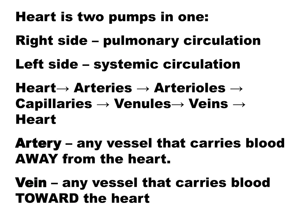
Heart is two pumps in one: Right side – pulmonary circulation Left side – systemic circulation Heart→ Arteries → Arterioles → Capillaries → Venules→ Veins → Heart Artery – any vessel that carries blood AWAY from the heart. Vein – any vessel that carries blood TOWARD the heart
Parietal pericardium: outer fibrous layer inner serous layer Pericardial cavity Visceral pericardium Pericarditis
Heart Wall • Layers (superficial to deep): • Epicardium – serous membrane • Myocardium – muscle layer • Endocardium – continuous throughout circulatory system
Magic School Bus https://www.youtube.com/watch?v=c7ulsNQzDxE https://www.youtube.com/watch?v=xDPpTVR3Q34
Cardiac Muscle: involuntary, striated Intercalated discs: gap junctions functional syncytium desmosomes – “spot welds”
Ventricular Geometry LV RV
Common Disorders Ischemia – reduced blood flow Hypoxia – reduced oxygen supply Angina pectoris – “strangled chest” referred pain Myocardial infarction – death of an area of tissue due to interrupted blood flow (“Heart attack”) Congestive heart failure – heart is unable to supply bloodflow to body Fibrillation – uncoordinated, unsynchronized beating of heart, no net bloodflow
Cardiac cycle • One complete heart beat: • Systole (contraction) and • Diastole (relaxation) of both ventricles • Remember: Blood pressure = systole/diastole ≈ 120/80 normal average
“Heart beat” • http://www.youtube.com/watch?v=ZlB-915CfCg • http://www.youtube.com/watch?v=7eFn8Cgcx8g • http://www.youtube.com/watch?v=NYB-rJZQt4w&feature=fvw
- Know http://www.youtube.com/watch?v=12_nJamoyTk
Cardiac Muscle Contraction http://www.youtube.com/watch?v=IjU81a5TjZs; http://www.dnatube.com/video/317/Beating-Heart-Cell; http://www.youtube.com/watch?v=55tAIOcBg3w&feature=related; http://www.youtube.com/watch?v=_gbGA5il4Sg • Heart muscle: • Is stimulated by nerves and is self-excitable (automaticity) • Contracts as a unit • Has a long (250 ms) absolute refractory period • Cardiac muscle contraction is similar to skeletal muscle contraction
Heart Physiology: Intrinsic Conduction • Autorhythmic cells: • Initiate action potentials • Have unstable resting potentials called pacemaker potentials • Use calcium influx (rather than sodium) for rising phase of the action potential http://www.youtube.com/watch?v=Lhl897Mz-h8&feature=related; http://www.interactivephysiology.com/demo/systems/buildframes.html?cardio/actnpot/01
Heart Physiology: Sequence of Excitation - know • Sinoatrial (SA) node generates impulses about 75 times/minute • Atrioventricular (AV) node delays the impulse approximately 0.1 second • Impulse passes from atria to ventricles via the atrioventricular bundle (bundle of His) http://www.smm.org/heart/heart/pumping.htm; http://depts.washington.edu/physdx/heart/demo.html
Heart Physiology: Sequence of Excitation - know • AV bundle splits into two pathways in the interventricular septum (bundle branches) • Bundle branches carry the impulse toward the apex of the heart • Purkinje fibers carry the impulse to the heart apex and ventricular walls
Extrinsic Innervation of the Heart - know • Heart is stimulated by the sympathetic cardioacceleratory center • Heart is inhibited by the parasympathetic cardioinhibitory center
Electrocardiography • Electrical activity is recorded by electrocardiogram (ECG) • P wave corresponds to depolarization of SA node • QRS complex corresponds to ventricular depolarization • T wave corresponds to ventricular repolarization • Atrial repolarization record is masked by the larger QRS complex http://www.youtube.com/watch?v=lYMSkGXFoN4&feature=related
Electrocardiography (EKG) http://www.youtube.com/watch?v=ew6Jp74vaN4
AED https://www.youtube.com/watch?v=ZRVFvtoLkRM
Heart Murmurs – abnormal sounds caused by the flow of blood. Mitral stenosis (abnormal narrowing) Mitral valve prolapse (turns “inside out”)
1. 2. 3.
Conduction system of the heart Sinoatrial (SA) node – “pacemaker’ → Atrioventricular (AV) node → Atrioventricular (AV) Bundle - Bundle of His→ Purkinje fibers – conduction myofibers Ectopic pacemaker = implanted device that uses electrical impulses to reproduce or regulate the rhythms of the heart
ECG (or EKG) • An electrocardiogram (ECG / EKG) is an electrical recording of the heart and is used in the investigation of heart disease. • Normal adult 12-lead ECG: