Download
1 / 33
330 likes | 350 Views
Explore the intricate anatomy of the ear, from the external to inner ear structures. Learn about sound transmission, auditory ossicles, and the physiology of hearing.

E N D
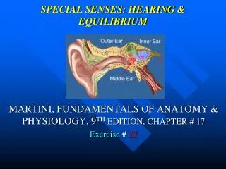
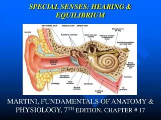
Parts of the Ear • external ear • middle ear • inner ear
Parts of the Ear inner ear external ear middle ear Eustachian tube
External Ear • collects sound waves and passes them inward into the external auditory canal • Includes: - auricle - external auditory canal - tympanic membrane
Auricle • flap of elastic cartilage, flared like a funnel • covered with thick skin • helix - top • lobule - bottom, earlobe • attached to head by ligaments and muscles
External Auditory Canal • curved tube about 1 inch long • skin-lined • near exterior opening are ceruminous glands - produce cerumen (wax) which helps to trap foreign material • lies in temporal bone • extends from auricle to tympanic membrane
Tympanic Membrane • thin, semitransparent membrane of fibrous connective tissue • lies between external auditory canal and middle ear • cone-shaped structure with apex directed medially • sound waves in auditory canal cause pressure changes that produce eardrum vibrations
Middle Ear • air-filled cavity in temporal bone • epithelium-lined • contains auditory ossicles • extends from eardrum to thin, bony partition with two membrane covered openings called oval and round window • connected to mouth by Eustachian tube
Auditory Ossicles • transfer vibrations from eardrum to oval window of internal ear • maleus is attached to the internal surface of eardrum at apex • incus is the intermediate bone • stapes is attached by ligaments to the membranous oval window
Auditory Ossicles (cont.) • malleous vibrates with tympanic membrane; passes vibration to incus • incus causes stapes to vibrate on oval window • oval window is pushed in and out, causing motion in fluid within internal ear • action activates receptor cells • impulses travel to temporal lobe of cerebrum and sound sensation results
Inner Ear • complex series of interconnecting chambers • Includes: - bony or osseous labyrinth - membranous labyrinth
Osseous Labyrinth • bony canal within the temporal bone • lined with periosteum • contains the fluid perilymph - chemically similar to cerebrospinal fluid • Divided into three areas: - semicircular canals - vestibule - cochlea
Membranous Labyrinth • surrounded by cerebrospinal fluid • series of sacs following general shape of osseous labyrinth • lined with epithelium • contains endolymph - chemically similar to intracellular fluid • portions within bony canals called semicircular ducts communicate with utricle and vestibule
Semicircular Canals • three canals; anterior, posterior, and lateral • each end enlarges into swelling called ampula • lie at right angles to each other • contain receptors for equilibrium
Vestibule • oval central portion of bony labyrinth • contains two sacs called the utricle and saccule - connect to each other by small duct • contains receptors for equilibrium
Cochlea (koklea = snail shell) • coil-shaped cavity • anterior to vestibule • makes almost three turns • central bony core called modiolus • contains thin, bony shelf which divides cochlea into upper and lower compartments and smaller cochlear duct • contains receptors for hearing
Three Compartments of Cochlea • scala vestibuli • scala tympani • cochlear duct (scala media)
Scala Vestibuli • above bony partition • ends at oval window • filled with perilymph Scala Tympani • below bony partition • ends at round window • contains perilymph
Cochlear Duct (scala media) • portion of membranous labyrinth • separated from scala vestibuli by vestibular membrane • separated from scala tympani by basilar membrane • organ of Corti (spiral organ) located on basilar membrane within cochlear duct • tectoral membrane projects over and in contact with hair cells of spiral organ
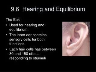
Organ of Corti • spiral organ • lined with epithelial cells - support cells and 16,000 hair cells contain receptors for auditory sensations
Hair Cells • Two types: - inner - outer • processes at apical end extend into endolymph of cochlear duct • synapse with fibers of the cochlear branch of the vestibulocochlear nerve (VIII) • easily damaged by high intensity sounds
Sound Waves • sound sensations are heard by vibrations transmitted through the air • result from alternate compression and decompression of air molecules • most audible sound vibrations to human ears are frequencies between 1000 and 4000 Hertz • entire audible range is 20 - 20,000 Hz
Pitch • determined by frequency of vibrations • the higher the frequency of vibrations, the higher the pitch (musical high note) Loudness • determined by amplitude of sound waves • measured in decibels (dB)
silence rustling leaves normal conversation crowd noise vacuum cleaner pneumatic drill uncomfortable sound painful sound 0 dB 15 dB 45 dB 60 dB 75 dB 90 dB 120 dB 140 dB Decibels of Sounds
Physiology of Hearing • auricle directs waves into external auditory canal • sound waves strike tympanic membrane • alternate compression and decompression of air cause membrane vibration • movement of membrane depends on intensity and frequency of sound waves • low-frequency = slow membrane vibration • high-frequency = rapid membrane vibration
Physiology of Hearing (cont.) • malleus connects in central area of tympanic membrane • malleus vibrates, conducting vibration to incus and then stapes • stapes pushes membrane on oval window in and out • movement of oval window causes waves in perilymph of cochlea
Physiology of Hearing (cont.) • inward movement of oval window pushes on perilymph of scala vestibuli to scala tympani to round window into middle ear • movement of perilymph exerts pressure on vestibular membrane • pressure in endolymph inside cochlea increases and decreases
Physiology of Hearing (cont.) • pressure fluctuations move basilar membrane causing hair cells of spiral organ to move against tectorial membrane leading to generation of nerve impulses in cochlear nerve fibers • pathway extends into medulla oblongata through midbrain to thalamus and on to temporal lobes of cerebrum for interpretation
Physiology of Equlibrium • when body movement occurs, organs detect motion and aid in maintaining balance • organs provide information on which way is up or down • Organs of equlibrium: - utricle - saccule - semicircular ducts
Two kinds of Equilibrium • static - maintenance of posture in response to changes in body orientation relative to the ground • dynamic - maintenance of body position, mainly the head, in response to sudden movements
Deafness • conduction deafness - impairment of structures that transmit vibrations - punctured eardrum, otitis media, wax buildup • nerve deafness - degeneration of receptors - damage to receptor cells
Ménière’s Syndrome • labyrinth disorder • characterized by fluctuating loss of hearing, vertigo, and tinnitus • caused by an increased volume of endolymph causing enlargement of the labyrinth • disease of cranial nerve VIII