Download
1 / 32
490 likes | 1.59k Views
Principles of MRI. Principles of MRI. Some terms: Nuclear Magnetic Resonance (NMR) quantum property of protons energy absorbed when precession frequency matches radio frequency Magnetic Resonance Imaging (MRI) uses spatial differences in resonance frequencies to form an image

E N D
Principles of MRI • Some terms: • Nuclear Magnetic Resonance (NMR) • quantum property of protons • energy absorbed when precession frequency matches radio frequency • Magnetic Resonance Imaging (MRI) • uses spatial differences in resonance frequencies to form an image • basis of anatomical MRI • functional Magnetic Resonance Imaging (fMRI) • exploits magnetic properties of hemaglobin to create images changes in cortical blood flow
Principles of MRI • Some terms: • Nuclear Magnetic Resonance (NMR) • quantum property of protons • energy absorbed when precession frequency matches radio frequency • Magnetic Resonance Imaging (MRI) • uses spatial differences in resonance frequencies to form an image • basis of anatomical MRI • functional Magnetic Resonance Imaging (fMRI) • exploits magnetic properties of hemaglobin to create images changes in cortical blood flow
Principles of MRI • Some terms: • Nuclear Magnetic Resonance (NMR) • quantum property of protons • energy absorbed when precession frequency matches radio frequency • Magnetic Resonance Imaging (MRI) • uses spatial differences in resonance frequencies to form an image • basis of anatomical MRI • functional Magnetic Resonance Imaging (fMRI) • exploits magnetic properties of hemaglobin to create images changes in cortical blood flow
Principles of MRI • Some terms: • Nuclear Magnetic Resonance (NMR) • quantum property of protons • energy absorbed when precession frequency matches radio frequency • Magnetic Resonance Imaging (MRI) • uses spatial differences in resonance frequencies to form an image • basis of anatomical MRI • functional Magnetic Resonance Imaging (fMRI) • exploits magnetic properties of hemaglobin to create images changes in cortical blood flow
Principles of NMR • Protons are like little magnets • they orient in magnetic fields like compass needles • what way do they normally point?
Principles of NMR • Protons are like little magnets • they orient in magnetic fields like compass needles • what way do they normally point? • normally aligned with Earth’s magnetic field
Principles of NMR • Protons are like little magnets • they orient in magnetic fields like compass needles • what way do they normally point? • normally aligned with Earth’s magnetic field • NMR uses a big magnet to align all the protons in a sample (e.g. brain tissue)
Principles of NMR • Protons are like little magnets • Radio Frequency pulse will knock protons at an angle relative to the magnetic field
Principles of NMR • Protons are like little magnets • Radio Frequency pulse will knock protons at an angle relative to the magnetic field • once out of alignment, the protons begin to precess
Principles of NMR • Protons are like little magnets • Radio Frequency pulse will knock protons at an angle relative to the magnetic field • once out of alignment, the protons begin to precess • protons gradually realign with field (relaxation)
Principles of NMR • Protons are like little magnets • Radio Frequency pulse will knock protons at an angle relative to the magnetic field • once out of alignment, the protons begin to precess • protons gradually realign with field (relaxation) • protons “echo” back the radio frequency that originally tipped them over • That radio “echo” forms the basis of the MRI image
Principles of NMR • Protons are like little magnets • The following simple equation explains MRI image formation
MRI Image Formation • First you need a scanner: • The first MRI scanner
MRI Image Formation • Modern Scanners
MRI Image Formation • Our Scanner
MRI Image Formation • Our Scanner
MRI Image Formation • Our Scanner
MRI Image Formation • Our Scanner
MRI Image Formation • MRI Image formation • resonance frequency depends on field strength • gradient coils alter resonance frequency over distance • slight differences in the “echo” frequency indicate the location of each proton • second-dimension of a slice is coded by the phase of the protons Increasing Field Strength field gradient = frequency gradient
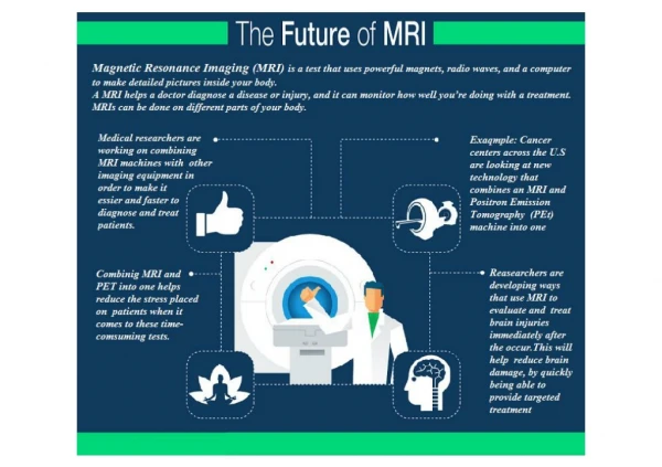
Functional Imaging • Functional Imaging must provide a spatial depiction of some process that is at least indirectly related to neural activity • in most imaging (i.e. PET, fMRI) that process is change in blood oxygenation related to changes in regional cerebral blood flow • Why should we measure blood oxygenation?
Functional Imaging • Why should we measure blood oxygenation? • Onset of a stimulus (or cognitive task) changes local blood oxygenation • first with a decrease • then with an “overshoot”
Functional Imaging • Why should we measure blood oxygenation? • Onset of a stimulus (or cognitive task) changes local blood oxygenation • first with a decrease • then with an “overshoot” • How do we measure changes in blood oxygenation?
Functional Imaging • Recall that precessing protons give off a radio “echo” as they realign with the magnetic field
Functional Imaging • Recall that precessing protons give off a radio “echo” as they realign with the magnetic field • We pick up the combined echo from many protons that are in phase
Functional Imaging • recall that the precession frequency depends on the field strength • anything that changes the field at one proton will cause it to de-phase
Functional Imaging • recall that the precession frequency depends on the field strength • anything that changes the field at one proton will cause it to de-phase • The de-phased region will give off less echo
Functional Imaging • Oxygenated hemoglobin is diamagnetic - it has no magnetic effects on surrounding molecules • Deoxygenated hemoglobin is paramagnetic - it has strong magnetic effects on surrounding molecules! Hemoglobin Heme
Functional Imaging • Oxygenated hemoglobin is diamagnetic - it has no magnetic effects on surrounding molecules • Deoxygenated hemoglobin is paramagnetic - it has strong magnetic effects on surrounding molecules! • Thus deoxygenated tissue gives of less MR echo because the protons de-phase quickly
Functional Imaging • blood flow overshoots baseline after a brain region is activated • More oxygenated blood in that region increases MR signal from that region (other regions de-phase faster)
Functional Imaging • It is important to recognize that fMRI “sees” changes in the ratio of oxygenated to deoxygenated blood - nothing more • BOLD: Blood Oxygenation Level Dependant contrast • How do we create those pretty pictures?