Download
1 / 68
1.79k likes | 5.09k Views
Wound Care. Jeanne Lowe PhD, RN, CWCN VA HSR&D Center of Excellence. Wound Care. Objectives: Describe skin function and structure Discuss normal phases of healing Identify factors that can interfere with normal healing Describe basics of wound assessment

E N D
Wound Care Jeanne Lowe PhD, RN, CWCN VA HSR&D Center of Excellence
Wound Care Objectives: Describe skin function and structure Discuss normal phases of healing Identify factors that can interfere with normal healing Describe basics of wound assessment Discuss different categories of wound dressings
Functions of the Skin • Protection • Thermoregulation • Sensation • Metabolism • Communication
Skin Structure Epidermis Dermis Subcutaneous Fat Muscle Bone
Factors Contributing to Impaired Skin Integrity • Circulation • Nutrition • Condition of the Epidermis • Allergies • Infections • Systemic Diseases • Trauma • Excessive Exposure • Mechanical Forces • Friction • Shearing • Pressure
Phases of Wound HealingHemostasis and Inflammation • Platelets release • vasoactive substance causing permeability • enzymes that attract leukocytes • growth hormones that influence fibroblasts • Wound develops erythema and edema
Phases of Wound HealingWound “clean up” • Neutrophils arrive • Phagocytosis • Macrophages appear within 3-4 days • Phagocytosis • Release of enzymes that trigger fibroblast response • Stimulate angiogenesis
Wound Repair • Regeneration of injured cells by cells of same type (i.e. Epidermis, bone) • Replacement by fibrous tissue (fibroplasia, scar formation)
Fibroplasia (Proliferation) • Occurs within the granulation tissue framework (new blood vessels and loose collagen) • Proliferation of fibroblasts at site of injury • Growth factors • Cytokines
Surgical Wound • Intentional injury that disrupts blood vessels and causes clotting and cascade of events that leads to wound closure within 2 to 4 weeks • History of Surgery • 18th Century surgeons were apprentices of barbers and butchers
Patient Risk Factors for Post-Surgical Wound Complications • Obesity • Diabetes • Immunosuppression • Cardiovascular disease • Smoking • Cancer • Previous surgery • Malnutrition
Surgical Wounds: Complications • Hemorrhage • Hematomas • Infection • Dehiscence • Evisceration • Fistula
Incision Healing Time • Epithelial resurfacing complete at 2-3 days • No tensile strength, but impenetrable to bacteria • “Healing ridge” 5-9 days • Lack of ridge = interventions to reduce incisional strain • Most dehiscences occur 5-8 days post-op, and about half are associated with infection
Incision Care • Cover with dry sterile dressing 24 to 48 hours, then open to air • Gently wash between sutures/staples to remove crusts • Report persistent pain, bleeding, erythema, wound edge separation or cloudy drainage
Wound Closure Aids • Steri-strip • Montgomery straps • Medical Staples • Sutures
Suture/Staple Removal • Usually removed 7-10 days post-op • Incisions over areas with tension up to two weeks • If concerned about incision dehiscence: • Remove every other one • Steri-strip
Wound Dehiscence • Fascial or Cutaneous disruption • Heavy bacterial load • Long time-lapse since wounding • Crushed or ischemic tissue – severe contused avulsion injury • Sustained high-level steroid therapy
Secondary Intention(includes chronic wounds) • Large tissue defect • More inflammation • More granulation tissue • Wound contraction - myofibroblasts
Factors Inhibiting Wound Healing • Medication • Cortisone, and epinephrine • Malnutrition • Protein & calories • Vitamin & mineral deficits • Zinc, Vitamin A, Vitamin C, Vitamin E • Dehydration • Edema • Perfusion • Chronic illness & other conditions • i.e. diabetes, CHF, immobility
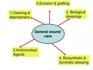
Principles of Wound Care • Keep wound moist • Manage drainage • Fill deep wounds • Control bacterial load • Protect wound from trauma • Assess healing
Keep Wounds Moist • Select dressings that maintain moisture. • Minimize time that wounds are open to air. • Add moisture to wound bed?
Manage Drainage • Maceration makes skin more fragile. • Excessive drainage requires nursing time.
Fill Dead Space • Fill wound with dressing • Be careful not to over-fill (no rocks)
Control Bacterial Load • Take time to wash or irrigate wounds to decrease bacterial load. • No need to scrub!
Protect From Trauma • Be gentle to skin • Use non-stick dressings • Minimize tape
But . . . • Remember to protect yourself from splash
Assess • Know what is under the dressing • Know typical healing pattern • Size matters • Document
Document findings • Location • Size (length / width / depth) • Wound base • Drainage • Surrounding skin • Systemic infection • What we’re doing
Wound Documentation:Wound Base Descriptors • Granulation tissue • Red, cobblestone/beefy. • Only in full thickness wounds • Epithelial tissue • Regrowth of epidermis • Pink or pearly • Smooth, shiny
Wound Documentation:Wound Base Descriptors • Slough • Necrotic/avascular tissue. • Moist. • Can be white, yellow, tan, or green. • Eschar • Necrotic/avascular. • Black or brown • Hard or soft. • Often leathery adherent tissue.
Wound Healing Basics • Wounds do best in moist environment • not too wet, not too dry • Loosely pack when needed • tight packing → injury to wound bed. • Protect peri-wound skin • No Sting Barrier • Cleanse/irrigate before assessment • Pre-medicate for pain prior to dressing changes • If culture is needed • cleanse wound thoroughly prior to swabbing • swab in area of granulation/viable tissue if present. • Never culture dressing!
Product Selection • Frequency of change • Ease of procedure • Caregiver ability • Availability of products • Cost/reimbursement factors
Dressing Purposes: • To absorb drainage • To prevent contamination • To prevent mechanical injury to the wound • To help maintain pressure to prevent excessive bleeding • To provide a moist wound environment • To provide comfort
Topical Wound Care Products • Alginates/Fiber Gelling Dressings • Antimicrobials • Collagen • Contact Layers • Foams • Gauze & Impregnated Gauze • Hydrocolloid • Hydrogels (Amorphous) • Skin Sealants • Topical Debriders • Negative Pressure Therapy • Compression Therapy
Gauze Packing(Kerlix, Nu-gauze, 4 x 4s) • description - inexpensive, user dependent • indications - to fill deep defects to maintain moisture and absorb exudate, may be soaked with antibiotic solution • considerations - pack lightly, may cause surrounding wound maceration, may traumatize wound if allowed to dry
Contact Layer Dressings (Greasy gauzes, N-terface, Adaptic, Xeroform, Mepitel) • description - nonadherent, prevents trauma and permits exudate to “pass through” pores of dressing for absorption by a secondary dressing, inexpensive • indications - superficial wounds with minimal to moderate exudate • contraindications - if goal is to “clean up” wound
Hydrocolloids(Duoderm, Comfeel) • description - absorbs exudate, maintains moisture, insulates, protects from secondary infection, non-permeable • indications - or superficial wounds with minimal to moderate drainage • contraindications - infected wounds Typically changed every 3 - 5 days
Polyurethane Foam (Mepilex,Biatain, LyoFoam) • description - nonadherent foam, absorbs exudate, insulates, variable protection from environmental contaminants (outer layer water proof or water-repellent) • indications - superficial weeping wounds, cover for deep (packed) wounds leave on for 3 - 5 days or change when cover-layer is at least 50% saturated