Download
1 / 50
570 likes | 1.3k Views
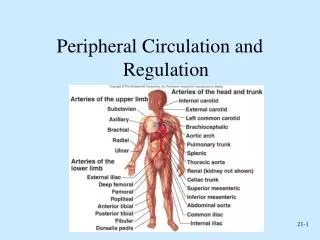
Blood, Blood Vessels & Circulation. Cardiovascular System. Blood vessels: Types. A. Arteries -carry oxygenated blood (most of the time) away from the heart -thicker than veins -three layers: inner endothelium middle smooth muscle outer connective tissue

E N D
Blood, Blood Vessels & Circulation
Cardiovascular System Blood vessels: Types A. Arteries -carry oxygenated blood (most of the time) away from the heart -thicker than veins -three layers: inner endothelium middle smooth muscle outer connective tissue -arteriole = small artery B. Veins -carry deoxygenated blood (most of the time) – toward the heart -same three layers as arteries (less SM and connective tissue) -thinner and more expansive than arteries -contain valves - to help the flow of blood back to heart -small vein = venule C. Capillaries -site of gas exchange with tissues -connect arterioles and venules -network of microscopic vessels (one cell thick) = capillary bed -site of exchange: gases, nutrients, wastes -can be closed off when not needed
Arteries • Tunica interna (intima) • simple squamous epithelium known as endothelium • basement membrane • internal elastic lamina • Tunica media • circular smooth muscle & elastic fibers • smooth muscle is innervated by sympathetic nervous system • decrease in stimulation or presence of certain chemicals causes vasodilation • increases diameter of vessel • nitric oxide, K+, H+ and lactic acid cause vasodilation • increase in stimulation causes muscle contraction or vasoconstriction • decreases diameter of vessel • Tunica externa • elastic & collagen fibers
Blood flow & Pressure gradients • blood flow is fastest in the arteries • slows within arterioles • slowest rate in capillaries - allows forexchange • blood flow becomes faster when vessels merge to form veins • flow rate through a vessel is proportional to the pressure gradient and inversely proportional to the vascular resistance (diameter) • F = ΔP/R • F = flow rate • ΔP = pressure gradient • R = resistance • ΔP is the pressure gradient = difference in P between the beginning and end of a vessel • this is what determines flow rate – NOT the absolute pressure! • resistance changes as the radius changes • R increases as r decreases – F will decrease (if P stays the same) • R decreases as r increases – F will increase (if P stays the same) • this relationship applies to non-viscous fluids
Blood flow & Pressure gradients • frictional losses (resistance) causes a drop in P as the blood travels through a section of vessel • caused by friction between the moving fluid and the stationary wall • if pressure gradient is unchanged – then increasing R will inhibit blood flow and decrease F • R depends on three factors • 1. blood viscosity (η) - # of circulating RBCs • 2. vessel length (L) • 3. vessel radius (r) – major determinant of R • R = 1/r4
Pouiseuille’s Law (Snooty French Guy Law) • -true description of how blood flow through our vessels – description of viscous fluids • Flow rate = η ΔP r4 • 8 ηL • -changing the viscosity (η) of the blood does NOT change the F value (go ahead try it!) • -but changing the L value does • -this is because thick, viscous blood “sticks” to the blood vessel wall • -so the longer the vessel – the more sticking and the slower the blood flows
Blood Pressure • Pressure exerted by blood on walls of a vessel • depends on the volume of blood within the vessel and the distensibility of the vessel • caused by contraction of the ventricles • highest in aorta • difference between systole and diastole – pulse pressure • the volume of blood entering an artery is not the same as the volume leaving it • during ventricular systole – the SV leaving the artery is 1/3 of that entering the artery • the artery will distend with increasing volumes • during diastole – the recoil of the vessel drives the exit of the blood • BUT no blood enters the vessel • mean arterial pressure – average pressure driving blood forward into the tissues throughout the cardiac cycle • at resting heart rate – about 2/3 of the cardiac cycle is spent in diastole • MAP = diastolic pressure + 1/3 (systolic pressure – diastolic pressure) • MAP falls steadily insystemic circulation with distance from left ventricle • 35 mm Hg entering the capillaries • 0 mm Hg entering the right atrium
mean arterial pressure – average pressure driving blood forward into the tissues throughout the cardiac cycle • at resting heart rate – about 2/3 of the cardiac cycle is spent in diastole • MAP = diastolic pressure + 1/3 (systolic – diastolic pressure) • MAP is what is regulated by homeostatic mechanisms • MAP falls steadily insystemic circulation with distance from left ventricle • 35 mm Hg entering the capillaries • 0 mm Hg entering the right atrium • our bodies need to control MAP – to keep it in a narrow range • we do this a few ways • 1. elastic arteries help decrease pressure in them by distending • 2. muscular arteries can vasodilate and decrease pressure in them by increasing diameter • 3. we can also decrease the SV of the heart
F = ΔP/R applies to the entire circulatory system in addition to a single vessel • F = cardiac output • ΔP = MAP (mean arterial pressure) • R = total peripheral resistance • total resistance offered by all systemic peripheral vessels • influenced by sympathetic nervous activity • rearrange the equation - ΔP = F X R • OR MAP = CO X total peripheral resistance
1 – CO and total peripheral resistance • 2 – CO depends on heart rate and SV • 3 & 4– heart rate depends on balance between the parasymp and symp divisions of the ANS • 5 – SV increases in response to symp activation (extrinsic control) • 6 – SV also increases with increasing venous return (instrinsic control) • 7- 10 – venous return is increased by symp induced vasoconstriction (7), skeletal muscle pump (8), respiratory pump (9) and cardiac suction (10) • 11 – 13 – venous return is also influenced by how much blood returns to the heart (11), actual blood volume (balance between passive bulk-flow fluid exchange between plasma and ECF (12), water and salt balance (13) and hormonal control (14)
15 & 16 – MAP is also determined by radius of the vessel (15) and the number of RBCs (16) • 17 - 20– arteriole radius can be controlled by metabolic factors which control blood need (17) - which leads to vasodilation (18) ALSO by sympathetic activity (19) which can cause vasocontriction (20) OR hormonally (20)
Arterioles • major resistance vessels in the vascular tree • radius is small enough to offer resistance to flow • high arteriolar resistance causes a marked drop in the MAP as blood flows through these vessels • MAP arteriole entrance = 93 mm Hg • MAP arteriole exit = 37 mm Hg • resistance also converts the pulsatile nature of systolic-diastolic pressure to nonpulsatile pressure within the capillaries • radius of the arteriole can be adjusted to • 1. variably distribute cardiac output among the organs • 2. help regulate arterial blood pressure
Arterioles and Arteriolar resistance • vasoconstriction and vasodilation • presence of a thick layer of smooth muscle • sensitive to many systemic, local factors + neurogenic stimulation • vascular tone • arterioles normally exhibit a state of slight constriction = vascular tone • helps establish a baseline of arteriolar resistance • tonic activity makes it possible to either vasoconstrict or vasodilate • two facets are responsible • 1. myogenic activity of smooth muscle layer • smooth muscle layer is responsive to neural or hormonal influences – self-induced contractile activity • 2. continuous release of NE by sympathetic fibers of the ANS
Arteriolar diameter • Intrinsic Factors/Local changes – at the tissue, organ specific • local chemical factors • metabolic factors • vasoactive mediators • histamine • local physical factors • hot/cold • passive stretch of arteriole • shear stress within arteriole • Extrinsic factors • sympathetic nervous system • hormones
Chemical influences and arteriole radius = local metabolic changes • most important local chemical influences on arteriolar smooth muscle are local changes in metabolism within that organ • local metabolic changes can affect the diameter of an arteriole without neural influence • active hyperemia = local arteriolar vasodilation that increases blood flow into an organ • arterioles are found within an organ and can be directly affected by that organ’s metabolic needs • local metabolic factors • decreased/increased oxygen = vasodilation/vasoconstriction • increase carbon dioxide = vasodilation • increased carbonic acid= vasodilation • increased K+ - repeated APs that outpace the Na/K pump’s ability to correct ionic changes = vasodilation • increased osmolarity – concentration of solutes accumulates in actively metabolic cells = vasodilation • prostaglandin release = vasodilation • relative concentration of these factors can determine the state of arteriolar muscle tone
Vasoactive Mediators • these local chemical changes do not act directly on smooth muscle but act on the endothelial cells • ECs – simple squamous epithelia cells • found lining the inside of the arteriole and capillary • ECs then release chemical factors called vasoactive mediators • e.g. endothelin = vasoconstriction • e.g. nitric oxide = vasodilation by relaxing arteriolar smooth muscle • inhibits entrance of calcium into the smooth muscle which inhibits the opening of the foot proteins on the sarcoplasmic reticulum • ECs have multiple roles
Histamine • NOT released by metabolic changes • NOT produce by endothelial cells • released upon pathology • released by connective tissue cells within the organ or by circulating white blood cells (mast cells, basophils) • usually released in response to organ damage • causes vasodilation to increase blood flow and speed healing
Local Physical factors • application of heat or cold • heat causes localized arteriolar vasodilation • increases blood flow • cold – counteracts histamine-induced swelling by inducing vasoconstriction • shear stress • blood flowing over the endothelial lining creates friction = shear stress • increase in shear stress can cause increased release of NO – promotes vasodilation • increased blood flow now reduces shear stress • myogenic response to stretch • increased MAP drives more blood into the arteriole which pushes out against the vessel wall = passive stretch • leads to vasoconstriction to reduce this blood volume • arteriolar smooth muscle responds to passive stretch by increasing its tone through vasoconstriction • this increased tone acts to resist the passive stretch • done by the release of vasoactive mediators from the EC (e.g. endothelin) • arterial occlusion can block blood flow and reduce this stretch • arterioles will dilate in response=reactive hyperemia
Extrinsic control of arteriolar diameter • includes both neural and hormonal control • sympathetic division innervates arteriolar smooth muscle everywhere except the brain • NO parasympathetic innervation of arteriolar smooth muscle! (exception – ciltoris and penis) • sympathetic activity contributes to arteriolar vascular tone • increased sympathetic activity induces vasodilation of arterioles in heart and skeletal muscle– drops arteriolar resistance and changes MAP • main area of the brain to adjust sympathetic output = cardiovascular control center • other brain regions involved – hypothalamus • Hormones: ADH, angiotensin II, epinephrine, norepinephrine
HORMONAL CONTROL OF ARTERIOLAR DIAMETER: • angiotensin II – converted from angiotensin I by the enzyme ACE (produced in the lungs) • regulates body’s salt balance • causes the release of aldosterone from the adrenal cortex – increased salt reabsorption • powerful vasoconstrictor • vasopressin (ADH) – released from the posterior pituitary in response to changes in water volume • drop in water content, release of vasopressin, decreased urine volume, increased water retention • also construction of peripheral vessels = vasoconstrictor • plays a role in reducing hemorrhage
-Epinephrine and Norepinephrine can be released from the adrenal medulla (sympathetic activity) or NE can be released from neurons (sympathetic) • -Ep and NE can cause vasoconstriction or vasodilation!!! depends on receptor and its location • -NE binds to a1-adrenergic receptors on ALL arteriolar smooth muscle to increase vasoconstriction • -Epi binds to b2-adrenergic receptors on arteriolar smooth muscle in heart and muscles to cause vasodilation • -cerebral arterioles do NOT have a1-adrenergic receptors !!! • -influenced entirely by local physical and chemical changes (intrinsic changes) • -during the “flight or fight” response - Epi is more abundant and has more affinity for the b2-adrenergic receptors which are expressed in large amounts on arteriolar smooth muscle in skeletal and cardiac muscle – so overall effect is vasodilation to heart and skeletal muscles
Evaluating Circulation • Pulse is a pressure wave • alternate expansion & recoil of elastic artery after each systole of the left ventricle • pulse rate is normally between 70-80 beats/min • tachycardia is rate over 100 beats/min/bradycardia under 60 • Measuring blood pressure with sphygmomanometer • Korotkoff sounds are heard while taking pressure • systolic blood pressure from ventricular contraction • diastolic blood pressure during ventricular contraction • provides information about systemic vascular resistance • pulse pressure is difference between systolic & diastolic • normal ratio is 3:2:1 -- systolic/diastolic/pulse pressure
the sympathetic nervous system essentially maintains an appropriate driving pressure to each organ (i.e. MAP) but the organ itself controls the amount of blood that actually enters it. • sympathetic control over arteriolar vascular tone constricts most vessels to ensure adequate MAP throughout the systemic circuit • BUT the organ can override the sympathetic control using local control mechanisms • analogy = water pressure in pipes
analogy – pipe carrying water • water pressure remains constant • differences in the amount of water entering the beaker depends on which valves are open and to what extent these valves are open • no water flows when the valve is closed • more water flows when the valve is wide open (low resistance) versus when a valve is partially open (high resistance) • so the sympathetic NS maintains the “water pressure” (i.e. the MAP), but each organ can control the amount of blood that enters them • when increased blood flows into one organ the others must compensate by changing their arteriolar diameter • SO THAT MAP IS MAINTAINED WITHIN AN APPROPRIATE RANGE
Regulation of BP • regulation of mean arterial pressure by the body done mainly through arteriolar diameter • Blood pressure = MAP within a small length of blood vessel • our body uses BP to immediately determine heart rate and contraction strength • BP is measured constantly by baroreceptors • changes in pressure within blood vessels • initiates either short-term or long-term reflexes • two major BRs: carotid sinus & aortic arch baroreceptors (mechanoreceptors) • as the BP within the vessels increase –increases the rate of firing of the afferent neurons within these BRs • decreasing pressure decreases the rate of firing • role of cardiovascular center in the medulla • help regulate heart rate & stroke volume • integrating center for information sent by the carotid and aortic BRs • divided into two centers: vasomotor & cardiac centers (made up of cardioacceleratory, cardioinhibitory divisions) • alters the ratio between sympathetic and parasympathetic activity to the heart and BVs • Vasomotor center = specific neurons regulate blood vessel diameter - sympathetic vasomotor nerves • Cardiac center = signals sent out through vagus & cardiac accelerator nerves - changes heart rate
Higher brain centers such as cerebral cortex, limbic system (emotions) & hypothalamus • anticipation of competition • increase in body temperature • Hypothalamus • osmoreceptors control salt and water balance and therefore long-term regulation of BP • cerebral cortex-hypothalamus preprograming – fight or flight responses, sexual orgasm, blushing • control over cutaneous arterioles for temperature regulation takes control over the cardiovascular center’s control over these vessels for regulation BP • therefore BP can fall dramatically as arteries dilate to allow cooling • Proprioceptors • input during physical activity • Chemoreceptors • monitor concentration of chemicals in the blood (O2, CO2 and H+ions) • also located in the carotid and aortic arteries • increase respiratory activity to bring in more O2 or blow off more CO2 • also increase BP by sneding excitatory impulses to the cardiovascular center
Continuous capillaries • skeletal & smooth, connective tissue and lungs • Fenestrated capillaries • plasma membranes have many holes • kidneys, small intestine, choroid plexuses, ciliary process & endocrine glands • Sinusoids • very large fenestrations • incomplete basement membrane • liver, bone marrow, spleen, anterior pituitary, & parathyroid gland Capillaries • Microscopic vessels that connect arterioles to venules • Found near every cell in the body but more extensive in highly active tissue (muscles, liver, kidneys & brain) • entire capillary bed fills with blood when tissue is active • lacking in epithelia, cornea and lens of eye & cartilage • Function is exchange of nutrients & wastes between blood and tissue fluid • Structure is single layer of simple squamous epithelium and its basement membrane
capillaries are not open under resting conditions in most tissues • prevents the flow of blood through the capillary bed • capillaries branch from a metarteriole or directly from an arteriole • metarteriole acts like a thoroughfare connecting a larger arteriole and a venule • these MAs are surrounded by spiralling smooth muscle cells – form a precapillary sphincter • the sphincters are not innervated by the nervous system by still possess a high degree of myogenic tone to contract in response to chemical factors released by the tissue • the more metabolically active the tissue, the greater the number of capillaries • e.g. muscle • only 10% of the capillaries are open in resting muscle • as the muscle increases its activity – local chemical factors change and the precapillary sphincters open to allow more blood flow
ECs fit together like a jigsaw puzzle with considerable gaps in between the cells = pores • sizes vary from capillary to capillary • brain capillaries have EC cells held together by tight junctions – no pores • most tissue capillaries allow the passage of small water-soluble substances like but glucose, small amino acids and peptides and ions • prevents the passage of larger proteins like plasma proteins • this transport may actually be regulated by the capillary itself • ECs may secrete substances that “tighten” up their junctions • histamine increases the gaps by inducing a contractile response in the EC and widening the gaps (actin-myosin interaction in the cytoskeleton) • in the liver, the capillary walls have larger pores to allow the passage of proteins • liver synthesizes the plasma proteins which must be allowed to pass into the circulatory system
Interstitial Fluid or ECF • passive intermediary between blood and cells – not a direct exchange between the blood and the ICF of cells • only 20% of the ECF circulates as plasma • the remaining 80% is interstitial fluid • exchange between the inside of the cell and the interstitial fluid (across the plasma membrane) is passive or active – depending on the cell and the solute being moved • BUT exchange between the interstitial fluid and the capillary wall is primarily PASSIVE • because the gaps in a capillary wall are quite large, the interstitial fluid and the composition of the blood are essentially the same • two ways to exchange materials between the blood and interstitial fluid • 1. diffusion • 2. bulk-transport
Capillary Exchange • Movement of materials in & out of a capillary • diffusion (most important method) • substances move down concentration gradient • all plasma solutes except large proteins pass freely across • through lipid bilayer, fenestrations or intercellular clefts • blood brain barrier does not allow diffusion of water-soluble materials (nonfenestrated epithelium with tight junctions) • transcytosis (vesicular transport) • passage of material across endothelium in tiny vesicles by endocytosis and exocytosis • large, lipid-insoluble molecules such as insulin or maternal antibodies passing through placental circulation to fetus • bulk flow • movement of large amount of dissolved or suspended material in same direction • move in response to pressure - from area of high pressure to area of low • faster rate of movement than diffusion or osmosis • regulates relative volumes of blood & interstitial fluid • comprised of: filtration (movement of material into interstitial fluid) + reabsorption (movement from interstitial fluid into capillaries)
Diffusion • diffusion is promoted by several factors: • 1. short distance of travel • 1. thinness of the capillary wall – 1 um in thickness (simple squamous epithelium + basement membrane) • 2. narrowness of the capillary – thinner than the RBC so the cells must pass through one at a time • 3. proximity to cells – every cell is approximately 0.01cm from a capillary • 2. total surface area – 10 to 40 billion capillaries • produces a tremendous surface area for exchange = 600m2 • 3. velocity of blood through the capillary • Slowest blood velocity is found in the capillaries • velocity is inversely proportional to the total cross-sectional area of all the capillaries • slow velocity of flow allows for sufficient exchange time • analogy – river – lake – river • flow rate (volume of water) flowing past any point along the river equals that of the lake • BUT the speed of the water flowing is slower in the lake
most capillary walls solutes cross via diffusion • so the chemical concentration of arterial blood is carefully regulated to promote the diffusion of specific solutes in a specific direction • the lungs add oxygen and remove carbon dioxide from the blood, the digestive organs supply glucose • the cells use this oxygen and glucose and replace them with waste products • the job of the blood is to continuously replace these waste products with fresh O2 and glucose • because O2 and glucose pass easily through the walls of the capillary – their movement is dependent upon their specific concentration gradient • as cells use more O2, this creates a larger concentration gradient between the cell and the blood – greater movement of O2
Bulk-flow • a volume of protein-free plasma filters out of the capillary and mixes with the surrounding interstitial fluid and then is reabsorbed = bulk flow • the various components are moved in bulk in contrast to the movement of individual components as seen in diffusion • when blood pressure inside the capillary exceeds that of the osmotic pressure of the blood – fluid is pushed out through the pores in the capillary wall = ultrafiltration • with the fluid comes the movement of multiple other components • when inward driving osmotic pressure exceeds the outward blood pressure– net inward movement of fluid and fluid components = reabsorption • ultrafiltration and reabsorption are collectively known as bulk flow • does NOT play a role in the exchange of individual solutes • does play a role in regulating the distribution of ECF between the circulating plasma and interstitial fluid • this is the way we establish the composition of our interstitial fluid
Bulk Flow • (Pc + πIF) – (πP + PIF) = outward pressure – inward pressure • major factors are Pc and πP
factors affecting bulk flow • 1. Pc = capillary blood pressure • fluid or hydrostatic pressure exerted on the inside of the capillary wall by blood • tends to force fluid OUT of the capillaries into the interstitial fluid • in general the blood pressure at the arteriolar end of a capillary = 37 mm Hg due to frictional losses in the upstream arterioles - declines to 17 mm Hg at the venous end of a capillary • 2. πP = plasma osmotic pressure (oncotic pressure) • osmotic pressure encourages inward movement of fluid into the capillary • 25 mm Hg – doesn’t change • 3. PIF = interstitial fluid hydrostatic pressure • fluid pressure exerted on the outside of the capillary wall by interstitial fluid • tends to force fluid into the capillaries and into the blood plasma • 4. πIF = interstitial fluid-colloid osmotic pressure (πIF is close to zero) • does not normally contribute significantly to bulk flow • Caused by the osmotic pressure of proteins in the IF - promotes the osmotic movement of water out of the blood and into the fluid • but during pathologic conditions – i.e. release of histamine which widens the capillary pores – increased leakage of plasma proteins into the interstitial fluid and πIF can increase
Net exchange of fluid across capillaries • Net exchange pressure = (Pc + πIF) – (πP + PIF) = outward pressure – inward pressure • two pressures forcing fluid out of the plasma are capillary blood pressure and interstitial fluid-osmotic pressure • two pressures forcing fluid into the plasma are plasma-osmotic pressure and interstitial fluid hydrostatic pressure • the imbalance between the forces produces a net exchange of fluid at a given point along the capillary wall • as the blood enters the capillary – outward forces exceed that of inward and fluid is forced out into the interstitial fluid (no protein movement!) – ultrafiltration forces result • at the venular end the inward pressure now exceeds the outward pressure and reabsorption will take place • as the blood moves through the capillary – pressure drops • so the amount of fluid moving out of the blood drops as the blood travels through the capillary region and progressively increasing amounts of fluid re-enter the blood as the blood reaches the venular end of the capillary • BUT these pressure values are controversial
Lymph • even under normal circumstances – more fluid is filtered from the blood plasma into the interstitial fluid than is reabsorbed back into the blood plasma • excess fluid = lymph • this excess fluid is directed into a series of vessels that are similar in structure and composition to veins – lymphatic vessels • one-way system of vessels that leads back to the circulatory system via the right lymphatic duct and the thoracic duct
Veins • Proportionally thinner walls than same diameter artery • tunica media less muscle – little inherent myogenic tone • lack external elastic lamina • tunica externa has more collagen than elastic fibers • strong and stretchy but possess poor elastic recoil • Still adaptable to variationsin volume & pressure • Valves are thin folds of tunica interna designed to prevent backflow • Venous sinus has no muscle at all • coronary sinus or dural venous sinuses
Blood Distribution • 60% of blood volume at rest is in systemic veins and venules • function as blood reservoir • often called capacitance vessels • venous capacity = volume of blood the veins can accomodate • body at rest – much of the blood bypasses the closed capillary beds and enters the venous return circuit – this increases the blood volume in the veins • 15% of blood volume in arteries & arterioles
Veins & Venules • veins & venules have little tone and resistance • the arteriole communicates chemically with the venule to ensure that inflow and outflow surrounding the capillary region matches • venous return is enhanced by many extrinsic factors • venous return = volume of blood entering the atria from the veins (pulmonary and systemic) • much of the driving force on the blood (blood pressure) has been lost as it enters the veins • BP averages on 17 mm Hg in the veins • but because the pressure of the blood entering the right atrium in o mm Hg – there is still a small driving force that helps to return the blood through the veins • clinical note – if atrial pressure becomes pathologically elevated (e.g. leaky AV valves) – this pressure differential can be lost, reducing the venous return
sympathetic activity – venous smooth muscle has an abundant nervous innervation by the sympathetic nervous division • can produce a small amount of constriction in the veins increasing venous BP and increasing the forces driving blood back to the heart • this contraction also decreases the amount of blood that can be held in the veins (decreases venous capacity) by decreasing the diameter of the vein itself • so less blood remains in the veins but is returned faster to the heart • skeletal muscle activity – contraction of skeletal muscles can push on the vein walls, decreasing their size and decreasing their capacity • venous valves – venous vasoconstriction and skeletal muscle contraction also drive blood away from the heart -evolved extensions off the endothelium – valves -these valves can shut off sections of veins to prevent back-flow towards the feet when standing • 4. respiratory activity– pressure in the chest is 5 mm Hg less than atmospheric • blood flowing through the thoracic cavity on the way back to the heart is exposed to subatmospheric conditions • this creates a pressure differential between the pressure in the limb veins and those in the thoracic region veins – drives more blood from the limbs = respiratory pump
Veins and gravity - gravity – when lying down, forces of gravity are equally applied to all veins -when standing, vessels below the heart become more subject to gravitational forces -so the veins increase their volume to counteract the increased volume -this reduces the amount of blood returning to the heart – decreases CO -also increases the pressure in the capillaries – forces more fluid out into the tissues from the blood -compensatory mechanisms – fall in MAP triggers the sympathetic NS to induce venous constriction to drive some of the pooled blood up toward the heart + skeletal muscle tone will increase to help propel blood to the heart