Download
1 / 61
630 likes | 656 Views
Discover the intricacies of microvascular decompression surgery for trigeminal neuralgia, a rewarding neurosurgical procedure with minimal side effects. Learn about patient selection, surgical techniques, imaging, and post-operative care.

E N D
Microvascular Decompression Surgery for Trigeminal Neuralgia Pearls and Pitfalls Aaron Cohen-Gadol, MD, MSc
Disclosures • Aaron A. Cohen-Gadol • Research Grants from Anspach and Zeiss
“TN/MVD – Desirable Practice” • TN is relatively easy to diagnose • MVD is one of the most satisfying operations in neurosurgery • Prescient/pleasing anatomy • Relatively short procedure • Microsurgical environment • Patients do well and are thankful • TN is “one of the worst pains humans have been afflicted with” • Minimal side-effects
TN Pain • Isolated to the 3 divisions of the nerve • Convulsive, not crossing midline • Has oral and skin triggers • Normal neurologic exam • Responds to Tegretol & antiepileptic drugs • The character of the pain may change with the use of neuropathic pain medications and have more of a constant character • Detailed history is important • Patients with predominantly constant or burning pain or facial numbness are not good candidates
Classification of TN Burchiel, Neurosurg Focus 18:1-3, 2005
Treatment • Medication • Surgical • Physiologic • Microvascular decompression (MVD) • Ablative • Radiosurgery • Percutaneous stereotactic rhizotomy (PSR) • Glycerol injection • Balloon compression
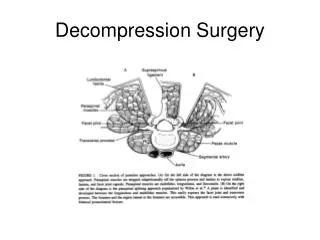
Pathogenesis and Treatment • No matter what the responsible etiology • MVD - Effective and durable palliative option • Percutaneous procedures - Less invasive • Posterior fossa exploration offers the only chance for a non-destructive procedure and a more durable result
Imaging for MVD • Brain MRI or CT scan • Exclude a structural pathology such as a meningioma, acoustic neuroma or an epidermoid tumor • High resolution MRI: Negative • Posterior fossa exploration is reasonable • We have routinely offered MVD to the patients who did not harbor an “MRI evident” vascular loop and have found compressive arterial loops during their posterior fossa exploratory surgery
Indications for MVD • Failure of medical or percutaneous therapy • Patient preference • Location of pain in V1 or multiple divisions • Physiologic young and can tolerate procedure • No perfect procedure, no cure • Be caring, listen and stay optimistic • Cure a few, comfort all! • Dysesthesia is not responsive to more procedures
Pearls • Control bleeding from the sinus with “ bone wax” and avoid aggressive packing • Control bleeding from SPV with gelfoam powder • Neurovascular conflict at the root entry zone of the nerve • Detailed and careful inspection of the space around the root entry zone (360) • Gentle handling of the nerve for a thorough inspection • The manipulation (“gentle” rhizotomy) of the nerve is responsible for some of the pain control afforded by MVD operations