Download
1 / 30
440 likes | 1.24k Views
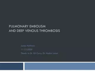
VENOUS THROMBOSIS. Dr Narisha Ramparsad Department of Haematology and Molecular Medicine. Normal haemostasis. Ensures fluid state of blood in vasculature Prevents blood loss – site of injury – by forming haemostatic plug Clot removal – when healing is complete. Overview of Haemostasis.

E N D
VENOUS THROMBOSIS Dr Narisha Ramparsad Department of Haematology and Molecular Medicine
Normal haemostasis • Ensures fluid state of blood in vasculature • Prevents blood loss – site of injury – by forming haemostatic plug • Clot removal – when healing is complete
Overview of Haemostasis THROMBOSIS BLEEDING
Definitions • A blood clot that forms in a blood vessel or within the heart and remains there is called a thrombus. A thrombus that travels from the blood vessel or heart to another location in the body is called an embolus, and the disorder, an embolism. For example, an embolus that occurs in the lungs is called a pulmonary embolism. • Sometimes, a piece of atherosclerotic plaque, small pieces of tumor, fat clumps, air, amniotic fluid, or other materials can act in the same manner as an embolus. (MEDLINE PLUS)
Pathogenesis -Thrombosis • Normally - balance between clotting and bleeding • With thrombosis - imbalance with procoagulant state manifesting • Risk increases when > 1 risk factor present • Venous thrombi – fibrin & RBCs mainly, leucocytes & plts also present • Virchow’s triad 1) vessel wall damage 2) blood flow (stasis) 3) hypercoaguability of blood Venous thrombosis can affect any part of venous system but deep veins most commonly affected.
Acquired Risk Factors • Malignancy • Presence of a central venous catheter • Surgery, especially orthopedic • Trauma • Pregnancy • Oral contraceptivesHormone replacement therapyTamoxifen • Immobilization • Congestive failure • Antiphospholipid antibody syndrome • Myeloproliferative disorders Polycythemia vera,Essential thrombocythemia • Paroxysmal nocturnal hemoglobinuria • Inflammatory bowel disease • Nephrotic syndrome • Hyperviscosity e.g Waldenstrom's macroglobulinemia, Multiple myeloma • Marked leukocytosis in acute leukemia • Sickle cell anemia
Acquired Risk Factors • NB in South Africa – effect of HIV • Decrease Protein S, Protein C • Increase Factor VIII
Inherited Risk Factors • Factor V Leiden mutation • Prothrombin gene mutation • Protein S deficiency • Protein C deficiency • Antithrombin (AT) deficiency • Rare disorders: Dysfibrinogenemia
Venous thromboembolism (VTE) • VTE = Deep Vein Thrombosis (DVT) and/or Pulmonary Embolism (PE) • Incidence increases with age • 117 cases/100 000 • Increasing health problem – prevention important, potentially fatal
VTE • Thrombosis commonly occurs in deep veins of limbs. Can also affect superficial veins • Venous system leg – 2 important categories 1- deep calf vein involvement only 2- proximal vein thrombosis (involving popliteal, femoral or iliac veins) – give rise to clinically significant PE • Pulmonary emboli ( majority arise from deep veins of leg >90%) • Other sources of PE include – upper extremity thrombosis, deep pelvic veins, renal veins, IVC
DEEP VEIN THROMBOSIS – clinical features • Leg pain, swelling, tenderness, palpable cord (thrombosed vessel), phlegmasia cerulea dolens in occasional patients. Non specific signs and symptoms.
Differential Diagnosis DVT • Cellulitis • Popliteal cyst • Lymphatic obstruction • Muscle strain/tear • Direct twisting injury to leg • If think about DVT – MUST objectively exclude
Pulmonary Embolism – Clinical Features • Symptoms variable • Transient Dyspnoea, tachypnoea • Tachycardia • Pleuritic chest pain, cough, haemoptysis, • CVS collapse with hypotension, syncope ( massive pulmonary embolism) • Clinically silent • Clinical features are non specific • ONCE again must objectively exclude
Laboratory investigations • D-Dimer assay • Compression ultrasound(DVT) • Venography (DVT) • Spiral CT (highly sensitive for PE • MRI • Work up for thrombophilic state when confirm diagnosis
Clinical course • Untreated proximal vein thrombosis – potentially lethal – fatal PE • Extension of thrombus proximally • Post thrombotic syndrome frequent complication of deep DVT. Heaviness, swelling cramps, itching, ulceration • Chronic thromboembolic pulmonary hypertension.
Treatment – Anticoagulant Therapy • Heparin: Unfractionated or Low Molecular Weight Heparins e.g Enoxaprin(Clexane), Dalteprin, • Fondaparinux • Vitamin K antagonists e.g Warfarin • Oral anti Xa (clinical trials) • Direct thrombin inhibitors
Initiation of treatment • Must cover with Clexane when initiating Rx with Warfarin – Why? ( short T1/2, Protein C) – relative prothrombotic state
Treatment – how long? • Individualise each case • Look at risk factors present • Transient vs permanent vs no risk factors • 1st episode vs recurrent thrombosis • Reassess • D-Dimer levels • Risk –Benefit ratio of anticoagulant therapy
Treatment – how long ? First onset ,transient risk factor – 6 months , recheck D-Dimer levels . If raised continue ? Indefinite therapy. First onset, idiopathic thrombosis – consider life long Warfarin. Reassess risk-benefit ratio Recurrent DVT – indefinite anticoagulation Patients with APL antibodies or 2 more inherited risk factors – 12 months anticoagulation and reassess
Treatment how long • First episode thombosis – with deficiency of natural anticoagulants e.g. antithrombin, Prot C, Prot S or FVLeiden/Prothrombin gene mutation – 12 months and reassess ? indefinite therapy
Side effects of anticoagulant therapy • Bleeding • Heparin Induced thrombocytopenia and thrombosis • Heparin induced osteoporosis, increased transaminase levels, hypersensitivity reactions e.g. necrosis, alopecia, hyperkalaemia
Treatment – Thrombolytic therapy – When? • Indicated in patients with PE – haemodynamically unstable , evidence of R ventricular failure • Threatened limb in setting of DVT
Prophylaxis – venous thrombosis • Important to identify those patient at increased risk of thrombosis • Prevention • Use of LMWH, compression stockings • Awareness – patients and health care professionals
CONCLUSION • Venous thromboembolism – major cause of morbidity and mortality • Identify risk factors early -institute prophylaxis • Prophylaxis imperative measure to decrease incidence of thrombotic events • If suspect thrombosis must objectively test • Duration of treatment – varies -individualise