Download
1 / 37
370 likes | 534 Views
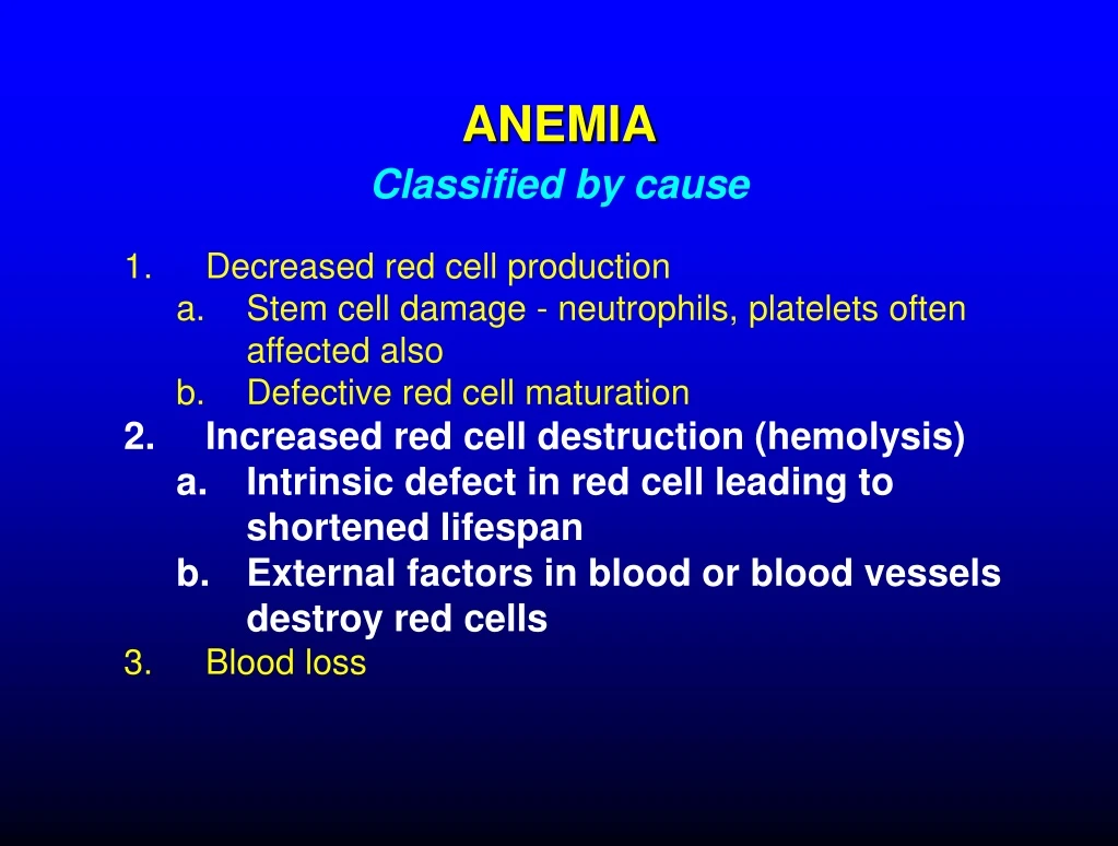
Classified by cause. ANEMIA. Decreased red cell production Stem cell damage - neutrophils, platelets often affected also Defective red cell maturation Increased red cell destruction (hemolysis) Intrinsic defect in red cell leading to shortened lifespan

E N D
Classified by cause ANEMIA Decreased red cell production Stem cell damage - neutrophils, platelets often affected also Defective red cell maturation Increased red cell destruction (hemolysis) Intrinsic defect in red cell leading to shortened lifespan External factors in blood or blood vessels destroy red cells Blood loss
HEMOLYTIC ANEMIA • Increased rate of red cell destruction • Increased rate of production - increased reticulocytes • Red cell destruction causes increased bilirubin production and jaundice • Most red cell destruction occurs in spleen • splenectomy may cause improvement
Hemolytic anemia: low hematocrit, plasma too yellow due to high bilirubin
INHERITED HEMOLYTIC ANEMIA Sickle cell anemia • Mutation changes structure of hemoglobin • Mutant hemoglobin (deoxy form) polymerizes in cells and damages cell membranes • membrane damage causes hemolysis • hemoglobin crystals change cell shape to "sickle" • sickled cells are rigid and block small blood vessels, causing tissue damage • Genetics: mainly affects those of African and Middle Eastern descent; recessive inheritance (carriers partially protected from malaria) O2 “Sickled” cell
SICKLE CELL ANEMIA Sickle cell Normal
Sickle Cell Normal red cell Sickle cells inflexible, can’t do this
IMMUNE HEMOLYTIC ANEMIA Production of "autoantibodies" against one's own red cells Antibodies coat cells and lead to destruction in spleen and liver Positive Coombs test (detects antibodies on red cells) in most cases Treatment: corticosteroids, splenectomy, i.v. gamma globulin
TRANSFUSION REACTION • Giving a person blood of the wrong type may cause destruction of the transfused cells (hemolysis) by antibodies in the recipient's blood • The most serious reactions occur with blood mismatched for antigens in the ABO system: • giving O patient A, B, or AB blood • giving A patient B or AB blood • giving B patient A or AB blood • In such instances there may be very rapid hemolysis accompanied by shock, kidney failure, bleeding, and death
HEMOLYTIC DISEASE OF THE NEWBORN • Caused by maternal antibodies against antigens on fetal red cells (usually Rh antigens); mother usually exposed (sensitized) to Rh antigen during prior pregnancy • These antibodies cross the placenta and cause destruction of fetal red cells • Infant liver unable to properly metabolize hemoglobin breakdown products (bilirubin) • Stillbirth or anemia, jaundice, and brain damage may result • Prevention: prevent sensitization by giving antibody against Rh factor (Rhogam) to Rh-negative woman soon after delivery of Rh-positive child
POLYCYTHEMIA • Definition: increased total red cell volume • high hematocrit • thick blood can cause thrombosis, other circulatory disorders • Polycythemia vera: increased, unregulated red cell production • Most cases due to an acquired mutation in marrow cells that makes red cell precursors much more sensitive to erythropoietin • Secondary polycythemia: increased erythropoietin production due to decreased oxygen delivery to kidney • Often due to low levels of oxygen in the blood
NEUTROPHIL DISORDERS • Neutropenia (decreased neutrophils) • Decreased production (bone marrow failure, cancer chemotherapy) • Increased consumption (some infections, enlarged spleen, autoimmune) • Increased risk of infection when neutrophil count low • Neutrophilia (increased neutrophils) • Increased production due to physiologic stimuli (e.g., infection) • Increased production due to bone marrow neoplasm
10/31/97 11/7/97 2/12/98 neutrophils 0 neutrophils 19,000 neutrophils 1200
LEUKEMIA • Malignant proliferation of white cells and/or their precursors (blasts) • Myelogenous (neutrophil precursors) • Acute myelogenous leukemia (AML) • Chronic myelogenous leukemia (CML) • Lymphocytic • Acute lymphocytic leukemia (ALL) • Chronic lymphocytic leukemia (CLL) • Chronic leukemias: more mature cells, slow-growing • Acute leukemias: immature cells (blasts), fast-growing
PATHOPHYSIOLOGY OF LEUKEMIA • Bone marrow failure (marrow fills with leukemic cells) • anemia • neutropenia (infections) • thrombocytopenia (bleeding) • Leukemic cells in blood may impair circulation • Leukemic cells in other organs • spleen, lymph nodes • skin • brain • Toxic substances from leukemic cells • uric acid (gout, kidney failure) • proteolytic enzymes (tissue damage, bleeding)
White cells Leukemia
ACUTE LEUKEMIAS • Acute myelogenous leukemia (AML) • adults > children • fatal if untreated • remission, occasional cure possible with intensive chemotherapy • sometimes curable with bone marrow transplant • Acute lymphocytic leukemia (ALL) • children and adults (most common childhood leukemia) • fatal if untreated • curable with chemotherapy or bone marrow transplantation • Cure rates in children > 75%
DIFFERENTIATION OF NEUTROPHILS AND RED CELLS Cells capable of division Cells cannot divide
Bone marrow in acute leukemia Normal AML
Immature cells (blasts) in acute myelogenous leukemia Mature lymphocytes in chronic lymphocytic leukemia
CHRONIC LEUKEMIAS • Chronic myelogenous leukemia (CML) • rare in children • treatable but often fatal within 5-10 years • Newer treatments will probably improve the prognosis • may be curable with bone marrow transplantation • Chronic lymphocytic leukemia (CLL) • almost all patients middle-aged and older • treatable but incurable • not all patients need treatment, many live > 10 years
Enlarged lymph nodes (lymphadenopathy) in chronic lymphocytic leukemia
LYMPHOMAS • Cancer of lymphocytes or their precursors • Forms tumors in lymph nodes, spleen, bone marrow, other organs • Sometimes in blood - overlap with lymphoid leukemia • Many different kinds - spectrum of severity • Hodgkin's vs non-Hodgkin's • B-cell vs T-cell • Mature vs immature lymphoid cells • Fast vs slow-growing • Usually treatable, sometimes curable (chemotherapy, radiation, marrow transplantation)
Many types, complex classification scheme NON-HODGKIN'S LYMPHOMAS LOW GRADE Cells appear mature Good news: slow-growing - patients may live many years with disease Bad news: treatable but generally incurable Usually affect older people HIGH GRADE Cells less mature The most aggressive forms resemble acute lymphoblastic leukemia Good news: often curable with chemotherapy Bad news: faster-growing, fatal in months if not treated or treatment doesn't work Some varieties occur in children Example: Burkitt lymphoma (resembles acute leukemia) Rule of thumb: the less mature the cancer cell, the faster growing the lymphoma, and the more likely the disease is to affect younger people and to be curable
HODGKIN'S DISEASE • Probably a cancer of lymphocytes (lymphoma) • Almost always begins in lymph nodes • Spreads gradually to other lymph nodes & organs • stage = extent of spread • Relatively common in young adults • Curable in many cases with radiation or chemotherapy “Reed-Sternberg” cell characteristic of Hodgkin’s disease
Staging HODGKIN’S DISEASE Stage I: single lymph node or contiguous group of nodes Stage II: more than one node group, same side of diaphragm Stage III: confined to nodes (and/or spleen) but present on both sides of diaphragm Stage IV: spread outside nodes (liver, bone marrow, lung, etc) Presence of symptoms (fever, weight loss, night sweats) designated by "B" after stage (no symptoms = "A") Lower stage disease often treated with radiotherapy; higher stage disease with chemotherapy
HODGKIN’S DISEASE Response to treatment Before treatment After 6 months (Treatment completed) After 7 years
MULTIPLE MYELOMA • Cancer of plasma cells (antibody-producing cells) • Most patients have monoclonal immunoglobulin (antibody-like protein) in blood or urine • This protein may damage kidneys, other organs • Bone destruction, bone marrow failure common
MULTIPLE MYELOMA Red cells stuck together by abnormal protein (rouleaux) Plasma cell