Download
1 / 34
380 likes | 450 Views
Explore the manifestations, diagnosis, and treatment of myocardial ischemia and heart failure, as well as the causes and symptoms of atherosclerosis and hypertension.

E N D
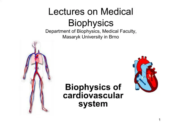
Cardiac Ischemia (myocardial ischemia) • Occurs when blood flow to your heart muscle is decreased by a partial or complete blockage of your heart's arteries (coronary arteries). The decrease in blood flow reduces your heart's oxygen supply. • It can damage your heart muscle, reducing its ability to pump efficiently. A sudden, severe blockage of a coronary artery can lead to a heart attack. • Causes: atherosclerosis of the coronary blood vessels blood clot
Manifestation of myocardial ischemia: • Chest pressure or pain, typically on the left side of the body • • Neck or jaw pain • • Shoulder or arm pain • • A fast heartbeat • • Shortness of breath when you are physically active • • Nausea and vomiting • • Sweating • • Fatigue
Manifestation of myocardial ischemia: • I-Angina pectoris: • An inadequate supply of oxygen to the heart muscle • *Is the major symptom of myocardial ischemia • *Presents as pain, pressure, or burning sensation in the area of the sternum, neck or jaw pain, shoulder or arm pain
Manifestation of myocardial ischemia: (cont.) II-Myocardial Infarction (Heart attack)A heart attack occurs when the flow of blood to the heart is blocked Cause of myocardial infarction: * Caused by occlusion of coronary blood vessels → death of myocardial tissue from ischemia and hypoxia * Most heart attacks results from occlusion of coronary BV by lipid deposits
*Manifestations of myocardial infarction: 1-Severe chest pain (pressing or crushing), accompanied by nausea, vomiting , sweating and weakness due to hypotension 2-Irreversible cell injury, occurs 20-30 minutes after complete ischemia 3-Release of myocardial enzymes: creatine phosphokinase (CPK) and lactate dehydrogenase (LDH) 4-ECG changes 5-Inflammatory response → leukocyte infiltration and fever.
Diagnosis of myocardial ischemia: 1- ECG (Electrocardiogram) 2- Stress test with ECG 3- Nuclear imaging 4- Cardiac catheterization Treatment of myocardial ischemia: Involves two strategies: A) Increase coronary blood flow by VD of coronary arteries B) Reduce cardiac work load by reducing heart rate and/or force of contraction
HEART FAILURE Is a condition in which the heart cannot pump blood effectively Left heart failure : There are two types of left-sided heart failure. •Systolic failure: The left ventricle loses its ability to contract normally. The heart can't pump with enough force to push enough blood into circulation. •Diastolic failure : The left ventricle loses its ability to relax normally (because the muscle has become stiff). The heart can't properly fill with blood during the resting period between each beat Fluid may back up in your lungs, causing shortness of breath -Result from diseases of myocardium and chronic hypertension -→Decreased stroke volume and Pulmonary congestion
Right heart failure : Occurs as a result of left-sided failure. When the left ventricle fails, increased fluid pressure is, in effect, transferred back through the lungs, ultimately damaging the heart's right side. When the right side loses pumping power, blood backs up in the body's veins. This usually causes swelling in the legs and ankles. -Occurs as a consequence of left heart failure or from chronic obstructive pulmonary disease, e.g, fibrosis
Atherosclerosis: Occurs when the arteries become thick and hard. is a condition in which plaque builds up inside the arteries. Plaque is made of cholesterol, fatty substances, cellular waste products, calcium and fibrin (a clotting material in the blood). Arteries contain an endothelium, a thin layer of cells that keeps the artery smooth and allows blood to flow easily. Atherosclerosis starts when the endothelium becomes damaged, allowing LDL cholesterol to accumulate in the artery wall.
Causes 1- High levels of cholesterol 2- Aging 3- Use of drug: e.g oral contraceptives. 4- Genetic Risk factors of atherosclerosis: 1-Smoking 2-Hypertension 3-Age < 45 years in males < 55 years in females 4-Elevated serum levels of LDL Low serum levels of HDL
Symptoms of atherosclerosis: Most symptoms of atherosclerosis don’t show until a blockage occurs. Common symptoms include: • chest pain or angina • pain in your leg, arm, and anywhere else that has a blocked artery • shortness of breath • fatigue • confusion, which occurs if the blockage affects circulation to your brain • muscle weakness in your legs from lack of circulation
Alteration of blood pressure Hypertension Consistent elevation of arterial pressure above the normal range expected for a particular age group Blood pressure is the force of blood against your artery walls as your heart pumps blood through your body. Hypertension occurs when the force of blood is stronger than it normally should be. For a patient to be diagnosed as hypertensive, he or she must have a blood pressure measurements of above 140mmHg orabove for systolic pressure and 90 mmHg or above for diastolic pressure Types of hypertension: • Primary or essential hypertension • Secondary hypertension • Malignant hypertension
I-Primary or essential hypertension • Most common type : 90-95% of hypertensive cases • Of unknown cause • Risk factors for developing primary or essential hypertension: 1-Familial history of hypertension 2-Increasing age 3-Race and gender: incidence is more in black men 4-High dietary salt intake 5-Obesity 6-Cigarette smoking . Manifestations of essential hypertension: *Most are asymptomatic *Frequent headaches
II-Secondary hypertension Some people have high blood pressure caused by an underlying condition. This type of high blood pressure, called secondary hypertension, tends to appear suddenly and cause higher blood pressure than does primary hypertension *Less frequent than essential hypertension (5-10%) *Causes: 1- Renal artery stenosis : narrowing of the arteries that carry blood to the kidneys narrowing caused by atherosclerosis of renal arteries. Decreased renal blood flow → increased *renin production → increased **angiotensin II formation → VC of blood vessels and salt and water retention → increased BP * an enzyme synthesized, stored, and secreted by kidney; it plays a role in increased of blood pressure **a vasoconstrictive substance formed in the blood when renin is released
2- Hyperaldosteronism: increased aldosterone secretion →salt and water retention → increased BP 3- Pheochromocytoma: (tumor of the adrenal gland) increased catecholamine secretion → VC of blood vessels + decreased urine formation → increased BP 4-Neurogenic :nervousness and anxiety → increased sympathetic discharge → VC of blood vessels → increased BP *Treatment of secondary hypertension: treatment of the cause
III- Malignant hypertension • Means dramatic increase of blood pressure (greater than 120-130 mmHg diastolic pressure) • It may occur suddenly in patients with essential hypertension • Is dangerous because it may damage : kidneys, retina, brain (edema and stroke) • Requires immediate medical treatment with powerful intravenous VD
Hypotension • Means abnormal low blood pressure • Most common form is orthostatic hypotension ( or postural hypotension) is an abnormal decrease in blood pressure when a person stands up • Causes of orthostatic hypotension : 1-Neurogenic: decreased nervous reflexes as occurs in old age 2-Decreased blood volume: caused by dehydration and diarrhea 3-Prolonged bed rest 4-Drug induced: use of antihypertensive drugs 5-idiopathic: cause is not known
Manifestations of orthostatic hypotension : • Dizziness • Decreased cardiac output • Decreased brain blood flow • Pooling of blood in extremities • Falls and injuries particularly in old age Treatment : 1-Maintains fluid volume 2-If patient is lying down, he should sit first for several minutes ,then stands slowly 3-Elastic stockings that prevents pooling of blood in lower limbs
Capillary circulation - Edema • The capillaries contain only 5% of circulating blood. This small volume of blood is the most important, because only in the capillaries the vital exchange of materials between blood and tissues takes place. • Exchange of materials across the capillary wall: Occurs mainly by filtration (A)The filtering force, that tends to move fluid outward is the hydrostatic capillary pressure (HCP), which is : At the arterial end=35 mmHg. At the venous end=15 mmHg.
Capillary circulation (contin) (B)The force that opposes filtration , that tends to move fluid inward is the colloid osmotic pressure of plasma proteins COP) which is about 25 mmHg. • So At the arterial ends of capillaries: Filtering force (HCP) 35mmHg Force opposes filtration (COP) 25mmHg Net filtering force = 10 mmHg • At the venous ends of capillaries: Filtering force (HCP) 15mmHg Force opposes filtration (COP) 25mmHg Net reabsorbing force = 10 mmHg
Capillary circulation (contin) • This means that the fluid filtered at the arterial ends = amount reabsorbed at the venous ends. • However, the amount filtered slightly exceeds that reabsorbed . This excess fluid returns back to the circulation through the lymphatic vessels.
Capillary circulation (contin) Edema • Definition: excessive accumulation of fluid in the interstitial spaces. • Edema occurs when tiny blood vessels in your body (capillaries) leak fluid. The fluid builds up in surrounding tissues, leading to swelling. • Types: generalized or localized
Causes: 1) Increased hydrostatic capillary pressure: as in a) Cardiac edema b) Pregnancy edema due to compression by pregnant uterus). c) Deep venous thrombosis.
Capillary Circulation Edema (contin) 2)Decreased colloid osmotic pressure of plasma proteins: caused by: a)Nutritional edema due to decreased protein intake in the diet. b)Hepatic edema caused by decreased synthesis of plasma proteins. c)Renal edema: caused by increased loss of plasma proteins in urine. 3)Increased capillary permeability: as in a) Allergy b) Inflammation 4) Lymphatic obstruction by filarial worms, e.g., elephantiasis.
Pathophysiology of veins Varicose veins: *Are veins that become distended over time due to pooling of blood in the lower limbs. That's because standing and walking upright increases the pressure in the veins of your lower body. • If the valves weaken or are damaged, the blood can flow backwards and collect in the vein, eventually causing it to be swollen and enlarged (varicose).
Manifestations of varicose veins 1-Aching pain 2-edema 3-Unsightly 4. dry skin and colour changes in the lower leg Treatment of varicose veins 1-Use of support stocking to prevent venous pooling 2- Surgical interventions may be used
Venous thrombosis: • Definition:formation of a blood clot in the lumen of veins • Predisposing factors: 1- Stasis of blood . 2-Hypercoagulability of blood. 3-Damage of blood vessels caused by trauma, surgery, catheters
Site of a thrombus: *Thrombi may form in : superficial vessels of the skin and extremities (benign and self limiting) or in deep veins →deep venous thrombosis , DVT (much more dangerous and require treatment) *Manifestations of DVT: 50% are asymptomatic Or it may present with pain, tenderness and swelling or embolism
Embolism: is a thrombus that breaks loose and travels through circulation Common sites of lodging of emboli: pulmonary, cerebral, and coronary blood vessels Embolism → ischemia and possible death of tissues due to blocking of blood flow Treatment and prevention of venous thrombosis: 1- Prevention of blood stasis: through ambulation, exercise and elevation of legs 2-Anticoagulant therapy e.g, warfarin ,heparin and aspirin 3-Thrombolytic therapy to dissolve the clot, e.g, streptokinase, tissue plasminogen activator (TPA) 4-Surgical removal of clots