Download

1 / 63
720 likes | 1.46k Views
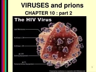
Characterizing and Classifying Viruses, Viroids, and Prions. 13. Characteristics of Viruses. Viruses Minuscule, acellular, infectious agent having either DNA or RNA Cause infections of humans, animals, plants, and bacteria Cause most of the diseases that plague the industrialized world

E N D
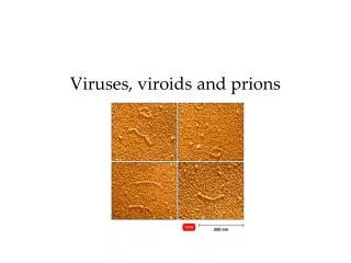
Characterizing and Classifying Viruses, Viroids, and Prions 13
Characteristics of Viruses • Viruses • Minuscule, acellular, infectious agent having either DNA or RNA • Cause infections of humans, animals, plants, and bacteria • Cause most of the diseases that plague the industrialized world • Cannot carry out any metabolic pathway • Neither grow nor respond to the environment • Cannot reproduce independently • Recruit the cell's metabolic pathways to increase their numbers • No cytoplasmic membrane, cytosol, organelles • Have extracellular and intracellular state
Characteristics of Viruses • Extracellular state • Called virion • Protein coat (capsid) surrounding nucleic acid • Nucleic acid and capsid also called nucleocapsid • Some have phospholipid envelope • Outermost layer provides protection and recognition sites for host cells • Intracellular state • Capsid removed • Virus exists as nucleic acid
Figure 13.1 Virions, complete virus particles, include a nucleic acid, a capsid, and in some cases an envelope. Capsid (sectioned to show interior) Nucleic acid (viral genome)
Characteristics of Viruses • Genetic Material of Viruses • Show more variety in nature of their genomes than do cells • Primary way scientists categorize and classify viruses • May be DNA or RNA, but never both • Can be dsDNA, ssDNA, dsRNA, ssRNA • May be linear and segmented or single and circular • Much smaller than genomes of cells
Figure 13.2 The relative sizes of genomes. Viral genome Partial genome of E. coli
Characteristics of Viruses • Hosts of Viruses • Most viruses infect only particular host's cells • Due to affinity of viral surface proteins for complementary proteins on host cell surface • May be so specific they only infect particular kind of cell in a particular host • Generalists – infect many kinds of cells in many different hosts
Figure 13.3 Some examples of plant, bacterial, and human hosts of viral infections.
Figure 13.4 Sizes of selected virions. E. coli (bacterium) (1000 nm x 3000 nm) Red blood cell (10,000 nm in diameter) Bacterial ribosomes (25 nm) Smallpox virus (200 nm x 300 nm) Poliovirus (30 nm) Bacteriophage T4 (50 nm x 225 nm) Bacteriophage MS2 (24 nm) Tobacco mosaic virus (15 nm x 300 nm)
Characteristics of Viruses • Capsid Morphology • Capsids • Provide protection for viral nucleic acid • Means of attachment to host's cells • Composed of proteinaceous subunits called capsomeres • Capsomere may be made of single or multiple types of proteins
Figure 13.6 The complex shape of bacteriophage T4. Head Tail fibers Tail Base plate
Characteristics of Viruses • The Viral Envelope • Acquired from host cell during viral replication or release • Envelope is portion of membrane system of host • Composed of phospholipid bilayer and proteins • Some proteins are virally coded glycoproteins (spikes) • Envelope proteins and glycoproteins often play role in host recognition
Figure 13.7 Enveloped virion. Glycoproteins Helical capsid Matrix protein Envelope Enveloped virus with helical capsid
Viral Replication • Dependent on hosts' organelles and enzymes to produce new virions • Lytic replication • Viral replication usually results in death and lysis of host cell • Five stages of lytic replication cycle • Attachment • Entry • Synthesis • Assembly • Release
Viral Replication: Overview PLAY Viral Replication: Overview
Figure 13.8 The lytic replication cycle in bacteriophages. Attachment Bacteriophage genome Entry Tail sheath Outer membrane Peptidoglycan Cytoplasmic membrane Bacterial chromosome Entry 2 Attachment 1 Phage DNA Lytic replication cycle of bacteriophage Bacterial chromosome degraded 3 Release 6 Synthesis 4 Phage proteins Assembly 5 Assembly Base Tail Mature head Tail fibers Sheath DNA Capsid Mature virion
Viral Replication: Virulent Bacteriophages PLAY Viral Replication: Virulent Bacteriophages
Viral Replication • Lysogeny • Modified replication cycle • Infected host cells grow and reproduce normally for generations before they lyse • Temperate phages • Prophages – inactive phages • Lysogenic conversion • Results when phages carry genes that alter phenotype of a bacterium
Figure 13.11 The lysogenic replication cycle in bacteriophages. Attachment 1 3 Prophage in chromosome Entry 2 Lambda phage Lytic cycle Lysogeny Synthesis 6 Release 8 4 Replication of chromosome and virus; cell division Assembly 7 Induction 5 Further replications and cell divisions
Viral Replication: Temperate Bacteriophages PLAY Viral Replication: Temperate Bacteriophages
Viral Replication • Replication of Animal Viruses • Same basic replication pathway as bacteriophages • Differences result from • Presence of envelope around some viruses • Eukaryotic nature of animal cells • Lack of cell wall in animal cells
Viral Replication • Replication of Animal Viruses • Attachment of animal viruses • Chemical attraction between viral protein and cell receptor • Animal viruses do not have tails or tail fibers • Have glycoprotein spikes or other attachment molecules that mediate attachment
Figure 13.12 Three mechanisms of entry of animal viruses. Phage genome inside capsid Cytoplasmic membrane of host engulfs virus (endocytosis) 1 2 3 Capsid 2 1 3 4 Receptors on cytoplasmic membrane Viral genome Direct penetration 6 5 Viral genome Viral glycoproteins remain in cytoplasmic membrane Viral glycoproteins 1 Uncoating capsid 2 Envelope 3 Endocytosis 4 Receptors on cytoplasmic membrane of host Viral genome Uncoating capsid Membrane fusion
Viral Replication • Replication of Animal Viruses • Synthesis of DNA viruses of animals • Each type of animal virus requires different strategy depending on its nucleic acid • DNA viruses often enter the nucleus • RNA viruses often replicate in the cytoplasm • Must consider • How mRNA is synthesized • What serves as template for nucleic acid replication
Viral Replication • Replication of Animal Viruses • Synthesis of DNA viruses of animals • dsDNA viruses • Similar to replication of cellular DNA • Viral genome replicated in the nucleus • Viral proteins are made in the cytoplasm • Some exceptions • Poxvirus replication occurs in the cytoplasm • Hepatitis B viruses replicate DNA from an RNA intermediary
Viral Replication • Replication of Animal Viruses • Synthesis of DNA viruses of animals • ssDNA viruses • Cells do not use ssDNA • Parvoviruses have ssDNA genomes • Host enzymes produce DNA strand complementary to viral genome to form dsDNA molecule • dsDNA used for viral replication and transcription
Figure 13.13 Synthesis of proteins and genomes in animal RNA viruses. –ssRNA virus Receptors on cytoplasmic membrane of host dsRNA virus +ssRNA virus +ssRNA –ssRNA dsRNA Transcription by viral RNA polymerase Transcription by RNA-dependent RNA transcriptase Unwinding –ssRNA +ssRNA acts as mRNA Complementary +ssRNA to act as template and as mRNA Translation of viral proteins, genome acts as mRNA Transcription by viral RNA polymerase to make complementary RNA strands Complementary –ssRNA to act as template Further transcription Further transcription Translation of viral proteins Copies of –ssRNA Translation of viral proteins Copies of +ssRNA Assembly Assembly Assembly Negative-sense ssRNA virus Double-stranded RNA virus Positive-sense ssRNA virus
Viral Replication • Replication of Animal Viruses • Synthesis of RNA viruses of animals • Retroviruses • Do not use their genomes as mRNA • Use DNA intermediary transcribed by viral reverse transcriptase as template to produce viral genomes
Viral Replication • Replication of Animal Viruses • Assembly and release of animal viruses • Most DNA viruses assemble in nucleus • Most RNA viruses develop solely in cytoplasm • Number of viruses produced depends on type of virus and size and initial health of host cell • Enveloped viruses cause persistent infections • Naked viruses are released by exocytosis or lysis
Figure 13.14: The process of budding in enveloped viruses. Enveloped virion 5 Budding of enveloped virus 4 3 2 1 Cytoplasmic membrane of host Viral glycoproteins Viral capsid
Figure 13.15 Pattern of virion abundance in persistent infections.
Viral Replication: Animal Viruses PLAY Viral Replication: Animal Viruses
Viral Replication • Replication of Animal Viruses • Latency of animal viruses • When animal viruses remain dormant in host cells • Viruses are called latent viruses or proviruses • May be prolonged for years with no viral activity • Some latent viruses do not become incorporated into host chromosome • Incorporation of provirus into host DNA is permanent
The Role of Viruses in Cancer • Cell division is under strict genetic control • Genes dictate that some cells can no longer divide at all • Cells that can divide are prevented from unlimited division • Genes for cell division "turned off" or genes inhibiting division "turned on" • Neoplasia • Uncontrolled cell division in multicellular animal • Mass of neoplastic cells is tumor • Benign vs. malignant tumors • Malignant tumors also called cancers • Metastasis occurs when tumors spread
Figure 13.16 The oncogene theory of the induction of cancer in humans.
The Role of Viruses in Cancer • Environmental factors that contribute to the activation of oncogenes • Ultraviolet light • Radiation • Carcinogens • Viruses
The Role of Viruses in Cancer • Viruses cause 20–25% of human cancers • Some carry copies of oncogenes as part of their genomes • Some promote oncogenes already present in host • Some interfere with tumor repression • Specific viruses are known to cause ~15% of human cancers • Burkitt's lymphoma • Hodgkin's disease • Kaposi's sarcoma • Cervical cancer
Culturing Viruses in the Laboratory • Viruses cannot grow in standard microbiological media • Cultured inside host cells • Three types of media for culturing viruses • Media consisting of mature organisms • Embryonated eggs • Cell cultures
Culturing Viruses in the Laboratory • Culturing Viruses in Mature Organisms • Culturing viruses in bacteria • Phages grown in bacteria in liquid cultures or on agar plates • Lysis of bacteria produces plaques • Allows estimation of phage numbers by plaque assay
Figure 13.17 Viral plaques in a lawn of bacterial growth on the surface of an agar plate. Bacterial lawn Viral plaques
Culturing Viruses in the Laboratory • Culturing Viruses in Mature Organisms • Culturing viruses in plants and animals • Numerous plants and animals have been used to culture viruses • Laboratory animals can be difficult and expensive to maintain • Ethical concerns
Culturing Viruses in the Laboratory • Culturing Viruses in Embryonated Chicken Eggs • Inexpensive • Among the largest of cells • Free of contaminating microbes • Contain a nourishing yolk • Fertilized chicken eggs are often used • Embryonic tissues provide ideal site for growing viruses • Some vaccines prepared in chicken cultures
Figure 13.18 Inoculation sites for the culture of viruses in embryonated chicken eggs. Air sac Injection into chorioallantoic membrane Injection into chorioallantois Injection into embryo Injection into amnion Injection into yolk sac