Download
1 / 99
1k likes | 1.22k Views
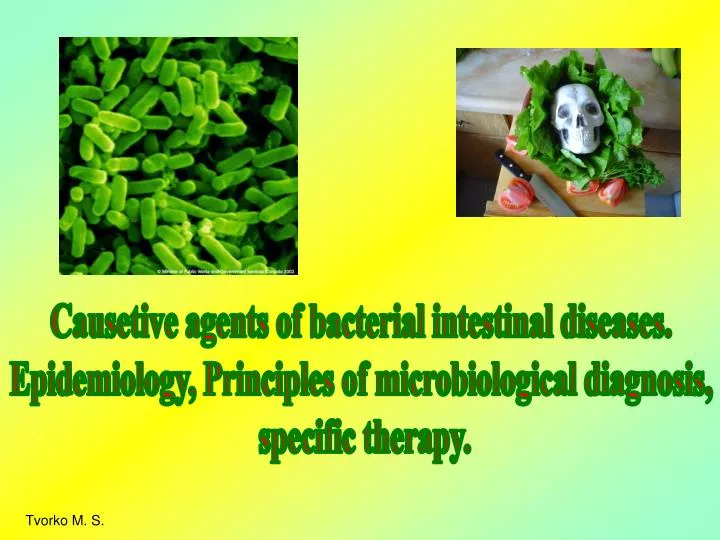
Causetive agents of bacterial intestinal diseases. Epidemiology, Principles of microbiological diagnosis, specific therapy. Tvorko M. S. Enterobacteriaceae. Escherichia coli Characteristics. Discovered in 19th century by Bavarian pediatrician Theodor Escherich

E N D
Causetive agents of bacterial intestinal diseases. Epidemiology, Principles of microbiological diagnosis, specific therapy. Tvorko M. S.
Escherichia coli Characteristics • Discovered in 19th century by Bavarian • pediatrician Theodor Escherich • Gram negative eubacteria • Facultative anaerobe • Usually motile • Universal inhabitant of human (mammalian) colon
Escherichia coli Found in the intestines of humans and animals, this bacterium is usually harmless, but some strains can cause food poisoning and more serious illnesses. Most outbreaks involve contaminated beef that was not cooked thoroughly. The strain known as O157:H7 is considered a potential biological weapon.
E. coli Lives in Colon of Healthy People (member of commensal flora) Includes E. coli
Cultivation. Colonies of E. coli on meat-peptone agar
Escherichia coli is highly motile and will show turbidity throughout the tube.
Fermentative properties. Positive (left) reactions of isolates E. coli in glucose fermentation broth. Note the formation of acid (yellow color) and gas. Observe the bubble in the Durham tube. “+” -vetest “—” -vetest
Indole reaction A.Salmonella B. E. coli is the positive microbe. A B
Note the bubble formation. Catalase positive E. coli can reduce nitrate to nitrite.
Antigenic structure. The antigenic structure of E. coli is characterized by variability and marked individuality. Along with the H- and O-antigens, the presence of other antigens has been shown in some strains, i.e. the surface somatic (membranous, capsular) K-antigens which contain the thermolabile L- and B-antigens and the thermostable A- and M-antigens. On the basis of antigenic structure an antigenic formula is derived which fully reflects the antigenic properties of the strain For example, one of the most widely spread serotypes is designated 0111 : K58 : H2.
Enteropathogenic E. coli (EPEC) • First E. coli pathotype described • UK pediatrician John Bray, 1945 • Causes potentially fatal infant diarrhea in developing areas • Contract organism by ingestion of contaminated water, food or fomites
EPEC Colonization and Lesion Development • patchy colonization of the small intestine • Generates unique histopathology termed “attaching and effacing” lesion • destroys microvilli • Expresses numerous toxins/effectors that manipulate host cell systems to serve the pathogen • EPEC, like other pathogenic E. coli strains, is a master cell biologist
Toxin production. • a gluco-lipo-protein complex with which their toxic, antigenic, and immunogenic properties • endotoxins • thermolabile neurotropic exotoxins • haemotoxins • pyrogenic substances, • proteinases, • deoxyribonucleases, urease, • phosphatase • hyaluronidase • aminoacid decarboxylases
Pathogenic E. coli Virulence Mechanisms Generalities • Virulence systems frequently encoded on mobile genetic elements • successful combinations reflected in pathotypes • Like other mucosal pathogens, pathogenic E. coli use multi-step strategy to infect host • attachment • evasion of host defenses • multiplication • damage host • Pathogenic E. coli oftencolonize host niches not normally inhabited by E. coli
Fig. 1.Scanning electron micrograph showing microcolonies of EPEC displaying the characteristic localized adherence pattern of adherence to HEp-2 cells. Fig. 2.High power scanning electron micrograph of EPEC displaying localized adherence to HEp-2 cells. Note the elongated microvilli to which the bacteria appear to attach.
Enterohaemorrhagic E. coli (EHEC) • First described in 1982 • Causes bloody diarrhea (haemorrhagic colitis), non-bloody diarrhea and haemolytic uremic syndrome (HUS) • ~5% of EHEC infections result in HUS; predominantly in children <5 years old and elderly • ~5% of HUS cases are fatal • Contract organism by ingesting contaminated food • common inhabitant of bovine gut • low infectious dose for humans (<100 organisms) • organism may be resistant to stomach acid
EHEC Virulence Factors • O157:H7 serotype dominant in North America, UK, Japan • O26 and O111 serogroups more prominent in other countries • Evolved from LEE containing EPEC serogroup O55 • Like EPEC, generates A/E lesion • Colonizes the large intestine • Expanded repertoire of adhesion factors • Encodes toxins/effectors in EPEC LEE • Additional virulence factors (plasmid and chromosomal) • RTX toxin similar to haemolysin • StcE activates host Complement cascade • Stx (Shiga) toxin
Stx (Shiga) Toxin • Toxin encoded on bacteriophage • >200 E. coli serotypes encode Stx • Shiga toxin E. coli (STEC) • most do not encode LEE (not virulent) • A/B toxin • pentameric B subunits bind holotoxin to host cell surface • A subunit cleaves host ribosomal RNA arresting protein synthesis and cell death (apoptosis) • Stx produced in colon travels through bloodstream to kidney • Damages renal endothelium and induces inflammation that may lead to acute renal failure and death
Shiga toxin • Distinguishing virulence factor • Subunit toxin: A: acts at ribosomal level, inhibits protein synthesis B: binds glycolipid receptor in mammalian cells (renal endothelium) • Stx1, Stx2 • Stx2 variants: 2c,2d,2e,2f
Excretion Re-colonization Environment Death Has HACCP led to a reduction in human incidence?
B.Enterotoxigenic E coli ( ETEC) • Common cause for travelers diarrhoea, and watery diarrhoea in children. • Colonisation factor facilitates the attachment to the intestinal epithelium. • Some ETEC produces heat labile exotoxin LT and heat stable or either of the toxins • LT has two sub units A B • Action -Activate Adenylate cyclase Increase local CAMP Intense, prolonged hypersecretion of water , Lumen of gut fill with water Hypermobility and diarrhoea results.
LT is antigenic and cross reacts with the enterotoxin of Vibrio cholerae. Some ETEC produces heat stable enterotoxin STa/b STa activates guanylyl cyclase. STb activates cyclic nucleotides. Releases water
Enteroinvasive E coli (EIEC) • Produces disease similar to shigellosis. • In adults this has been isolated with Shigella • Commonly affect children in developing countries, • and travelers. • Disease is due to invasion into mucosal cells of the intestine • multiply inside the cells and destruction /inflammation/ulceration • diarrhoea with blood • EIEC are nonlactose fermenter, or late lactose fermenter • and non motile.
E. coli Enteroinvasive E. coli • member normal gut flora • Agent of bacillary dysentery like disease
Normal Colon Dysenteric Colon • healthy GI tract • diseased GI tract • Fever • Severe abdominal pain • Bloody discharge with mucous
E. Enteroaggregative E coli (EAEC) • Produce acute/chronic diarrhoea in persons in developing countries. • Sepsis When normal host defense is poor sepsis can happen. • Common in new born babies whose IgM level is low.
Treatment of E.coli related diarrhoea 1st Line • Nitrofurantoin • Nalidixic acid • Norfloxacin ABST’ SHOULD BE DONE • Ampicillin • Cotrimoxazole 2nd line • Ciprofloxacin/Ceftriaxone/Cefuroxime Gentamicin
Salmonella • Salmonella enterica • one species, ~2000 serovars • Non standard nomenclature • S. enterica serovar Typhimurium • or S. typhimurium • rod-shaped, non-spore-forming Gram-negative bacterium • belongs to the family Enterobacteriaceae • close relative of E. coli • Motile by peritrichous flagella (H antigen). • nonmotile exceptions: S. gallinarum and S. pullorum
Enteric fever typhoid and paratyphoid fevers typhi, paratyphi A, B, C systemic infection infects only humans GI symptoms may not be evident Salmonella gastroenteritis non-typhi serovars zoonosis: predominantly food-borne can be complicated by septicaemia more common with some serovars, e.g. S. dublin (15% mortality rate when septicemic in the elderly) Metastatic disease, e.g. osteomyelitis Salmonella infections in humans
10 most frequently isolated Salmonella strains causing human disease • S typhimurium (22.1%) • S enteritidis (26.1%) • S enteritidis heidelberg (4.8%) • Salmonella enteritidis newport (4.3%) • Salmonella hadar (2.7%) • Salmonella enteritidis agona (2.0%) • Salmonella enteritidis montevideo (1.7%) • Salmonella oranienburg (1.6%) • Salmonella muenchen (1.5%) • Salmonella enteritidis thompson (1.5%)
Salmonella typhi Scanning electron micrograph Gram’s staining