Download
1 / 35
390 likes | 765 Views
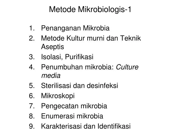
Metode Mikrobiologis -1. Penanganan Mikrobia Metode Kultur murni dan Teknik Aseptis Isolasi, Purifikasi Penumbuhan mikrobia: Culture media Sterilisasi dan desinfeksi Mikroskopi Pengecatan mikrobia Enumerasi mikrobia Karakterisasi dan Identifikasi. 1. Penanganan mikrobia.

E N D
Metode Mikrobiologis-1 Penanganan Mikrobia Metode Kultur murni dan Teknik Aseptis Isolasi, Purifikasi Penumbuhan mikrobia: Culture media Sterilisasi dan desinfeksi Mikroskopi Pengecatan mikrobia Enumerasi mikrobia Karakterisasi dan Identifikasi
1. Penanganan mikrobia Safe handling microbes: • Sebagian besar mikrobia bermanfaat • Sebagian kecil mikrobia merupakan agensia penyakit e.g. HIV, Neisseria meningitis • Kultur mikrobia tidak terlepas ke lingkungan • Kultur mikrobia tidak terkontaminasi oleh mikrobia lain • Teknik saftey harus dipenuhi dalam bekerja dengan mikrobia • Bahan dan alat dan tempat bekerja harus steril (teknik aseptis) • Kultur mikrobia yang tidak dipakai lagi harus dimusnahkan
2.Metode kultur murni (Pure culture) • Mikrobia yang dipelajari harus dalam bentuk kultur murni • Kultur murni: kumpulan sel yang merupakan keturunan sel tunggal • Strain : kumpulan sel kultur murni yang berasal dari satu sel tunggal • Untuk memperoleh kultur murni →Teknik Aseptis • Teknik Aseptis: semua bahan, alat, dan tempat bekerja yang digunakan serta cara kerja harus bebas dari kontaminasi(steril)
Kultur murni mikrobia Inokulasi Koloni
Aseptic Technique • A pure culture is a culture consisting of only one kind of microorganism • Aseptic technique is technique which involves avoiding any contact of the pure culture, using sterile medium, and using sterile surfaces of the growth vessel with contaminating microorganisms • How to accomplish it? • Sterilization of work area • Sterilization of working instruments and medium • Work must be done quickly and efficiently
3. Isolasi dan Purifikasi: Teknik Enrichment dan Isolasi Selektif
Enrichment Technique • Method used to isolate specific groups of microorganism • Culture medium and incubation conditions are designed to support target microorganism • Microorganisms other than the target will grow poorly or unable to grow in enrichment medium
Isolation of Microorganism • Isolation of pure cultures involves: • Separating samples of microorganisms into individual cells • Allowing the individual cells to reproduce and form clones of single microorganism • The success of isolation depends on: • Ability to do physical separation of the microorganism • Ability to maintain viability and growth of pure culture of microorganism
Isolation Diagram Samples Enrichment Dilution Platting Streak Plate Spread Plate Pour Plate Maintenance
4. Penumbuhan Mikrobia : Culture Media • A suitable culture media for microorganisms growth must: • Containing substances which support their needs. Substances (nutrients) divided into three types: macronutrients (C, N, P, S, K, Mg, Ca, Na, Fe), micronutrients(Cr, Co, Cu, Mn, Mo, Ni, Se, W, V, Zn), and growth factors (vitamins, amino acids, purines, pirimidines) • Having suitable environment conditions (pH, temperature, oxygen level)
Types of Culture Media • Defined media: media in which all components are known( eg. Azotobactermedium, BG-11 Medium) • Complex media: media in which the components are not totally known and may vary (eg. NA, PDA) • Selective media: media which only favor the growth of specific microorganisms (eg. Salmonella-Shigella Agar) • Differential media: media which contain substances that can detect microorganisms with specific metabolic activitise (eg, MacConkey Agar)
Maintenance and Preservation of Pure Cultures • Maintenance of microorganism is important to preserve cultures which have special features • The purposes of maintenance are to maintain viability and genetic stability of the microorganisms • Several methods of maintenance are: subculture, drying, freeze-drying, and freezing
5. Sterilisasi dan disinfeksi • Steril: bebas dari segala bentuk kehidupan • Sterilisasi: upaya untuk mencapai keadaan steril • Bactericidal • Fungicidal • Viricidal (seolah virus jasad hidup !) - inactivator • Germicides • Bacteristatic • Fungistatic • Antibiotics
5. Sterilisasi dan disinfeksi • Desinfection • Desinfectan • Antisepsis • Antiseptic • Sanitation
Metode Sterilisasi Metode Fisikawi: • Mekanis : filtrasi • Pemanasan: • Pemijaran: e.g. pemijaran ose • Udara panas kering: e.g. oven • Uap air panas: e.g. Arnold –steam sterilizer • Uap air panas bertekanan: e.g. Autoclave • Penyinaran : • Radiasi UV, gamma
Sterilization • Sterilization is a process to eliminate contaminant microorganisms • Chemical sterilization (alcohol, xylol, H2O2 etc) --> work surface and working instrument • Radiation sterilization (UV, gamma, etc) --> work surface and working instrument • Filtration --> heat-sensitive growth medium • Heating (autoclave, oven) --> working instrument, heat-resistant growth medium
Metode Sterilisasi Metode Kimiawi: • Disinfectant: HgCl2, betadine • Antiseptics: fenol, cresol • Antibiotics • Ozonization (ozon)
6. Mikroskopi Light microscopy: • Bright-field microscopy • Dark-ground microscopy • Phase-contrast microscopy • Fluorescence microscopy Electron microscopy: • Scanning Electron Microscopy (SEM) • Transmission Electron Microscopy (TEM)
Microscopy Technique • Microscopy is the use of microscope to view objects too small to be visible to naked eye • Type of microscope: • Light microscope (brightfield, darkfield, ultarviolet, fluorescence, phase contrast, nomarski differential interference, confocal) • Electron microscope (Transmission Electron Microscope, Scanning Electron Microscope)
Confocal microscopy • Confocal scanning laser microscopy (CSLM) • Memakai sinar LASER • Bayangan tiga dimensi dan lebih jelas Scaning Probe Microscopy • Scanning tunneling microscope (1980) • G. Binnig & H. Rohrer : Nobel Prize (1986) • Perbesaran 100 juta kali • Dapat melihat atom pada permukaan padat • Atomic force microscope (lebih baru) • Spesimen yang non-konduktor: interaksi protein, letak restriction site pada plasmid
7. Pengecatan mikrobia • Simple staining • Gram staining • Ziehl-Neelsen staining • Granules staining • Negative staining • Spore staining
Staining • Staining is important to discerns object from its background, hence it is become viewable under light microscope • Staining procedure: • Simple staining (positive staining, negative staining) • Differential staining (Gram staining, acid-fast staining, endospore staining)
Simple Staining • Single stain is used • All cells and structures generally stain the same color • Positive staining: stain attracted to the cell • Negative staining: stain repelled by the cell, background takes on the color
Differential Staining • Multiple staining reactions are employed • Differentiate types of cells or cell’s structures base on their staining reactions, hence given different color • The specimen must be “fixed” by heating or chemical • Examples: • Gram stain: staining based on composition of lipids and peptidoglycan in bacterial cell wall • Acid-fast stain: staining based on composition of mycolic acid, fatty acids, waxes, and complex lipids in bacterial cell wall • Endospore stain: staining to visualize bacterial endospore
Gram staining Acid-fast staining
Acid-fast Staining Gram Staining Endospore Staining