Download
1 / 1

E N D
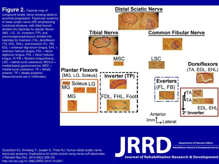
Figure 2. Fascicle map of compound sciatic nerve showing distal to proximal progression. Fascicular anatomy of lower sciatic nerve (2R) emphasizing functional divisions, with tibial branch divided into fascicles for plantar flexion (MG , LG , S), inversion (TP), and commonperoneal branch divided into fascicles for inversion (TA), dorsiflexion (TA, EDL, EHL), and eversion (FL, FB). EDL = extensor digi-torum longus, EHL = extensor hallucis longus, FDL = flexor digitorum longus, FHL = flexor hollucis longus, FL/FB = fibularis longus/brevis, LSC = lateral sural cutaneous, MG/LG = medial/lateral gastrocnemius, MSC = medial sural cutaneous, TA = tibialis anterior, TP = tibialis posterior. Measurements are in millimeters. Gustafson KJ, Grinberg Y, Joseph S, Triolo RJ. Human distal sciatic nerve fascicular anatomy: Implications for ankle control using nerve-cuff electrodes. J Rehabil Res Dev. 2012;49(2):309–22.http://dx.doi.org/10.1682/JRRD.2010.10.0201