Download
1 / 55
1.19k likes | 2.53k Views
Obstetric Ultrasound. Felipe Moretti, MD Griff Jones, MD, FRCS Assistant Professor – Uottawa Maternal Fetal Medicine Division.

E N D
Obstetric Ultrasound Felipe Moretti, MD Griff Jones, MD, FRCS Assistant Professor – Uottawa Maternal Fetal Medicine Division
“About 4% of all pregnancies are complicated by one or more major fetal malformations, 2% by a fetal genetic disorder, 1% by miscarriage after the first trimester, and another 1% result in infant death in the first year of life”. • Obstetrical and Gynecological Survey, 2008.
What is Prenatal Diagnosis? Aneuploidy Downs’ Syndrome Anomalies Spina bifida / NTD Fetal diseaseIso-immunisation (Rh) Infection Cardiac arrythmias
Obstetric Ultrasound First Trimester scan Second trimester Third trimester
Obstetric Ultrasound First Trimester scan Determine Gestational age Viability Number of embryos or fetus Intrauterine pregnancy
First Trimester scan Determine Gestational age: CRL (Crump Rump Length)
Normal Early Pregnancy Fetal cardiac activity present
First Trimester scan Number of embryos or fetus
First Trimester scan IPS ( Integrated Prenatal Screen): Combine test with Maternal blood work and Ultrasound Blood work and US at 11-13+6 days: pregnancy associate plasma protein-A (PAAP-A) and free-hCG plus NT Blood work at 15-19 weeks: AFP, estriol and inhibin.
Fetal Structural Anomalies • Anatomy review done at 18 - 20 weeks • Striking a balance • Adequate visualisation of fetal structures • Allow adequate time for further investigation • Leave parents the option of not continuing the pregnancy • Studies have shown a higher detection rate at 20+ weeks
Difficulties in Imaging Obesity
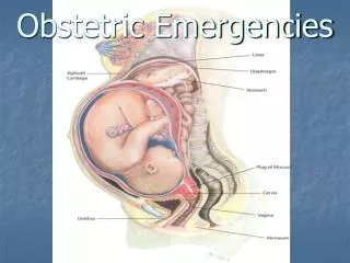
Fetal Position • Apposing structures • Shadowing • Orientation • TV scanning • Heart • Head • Engagement
Congenital Cardiac Anomalies • Detection rate remains poor • 25-50% • Technically difficult • Complex anatomy • Movement • Function changes at birth
Level 2 Ultrasound for Maternal Valproate Exposure “Normal” genetic sonogram “Normal” extended anatomy review Baby discovered to have Downs’ Syndrome at birth
Prenatal ultrasound is not a perfect science Risks are modified Nothing is 100%
What else Ultrasound can help us in the 2nd and 3rd trimester? Placenta Location; Anterior/Posterior/Fundal/Lateral Previa or non-previa Presentation; Cephalic Breech Transverse
Fetal Growth • There is a higher morbidity and mortality in babies that are small for gestational age • Unfortunately, most babies weighing <10th centile are “normally” grown and a significant number of IUGR babies have birthweights >10th centile
Fetal Measurement 2nd and 3rd trimester Fetal Head: BPD and HC Fetal Abdomen: AC Femur Length Estimate Fetal Weigth (EFW)
Variability in Weight Estimates • Technical / image quality • Caliper placement • Numerous mathematical models • Log weight = 3-1.7492+0.0166(BPD + 0.0046*AC – 0.00002646*AC*BPD) • All tend to be poor at weight extremes • Aim for +/- 10% in 90% estimates
Ultrasound Assessment of Fetal Behaviour • Significant Canadian contribution to the field • Followed on from the introduction of real-time ultrasound • Led to the development of the Biophysical Profile (BPP)
Fetal Breathing • Occurs 30% of time at term • Clusters lasting 20+ minutes every 60-90 minutes • Apnea episodes lasting up to 2 hours occasionally seen
Fetal Movement • Fetus moves 10% of time at term • Average of 31 movements per hour • No movement occasionally occurs for up to 75 minutes
Fetal Tone • One episode of extension and return to flexion in 30 min • More recent modification to reflect fine motor activity • Hand opening / closing • Mouth opening / closing
Component Criteria to score “2” 1.Breathing movements At least one episode continuing more than 30 seconds. Hiccoughs / hiccups count. 2.Body Movements At least three body or limb movements. 3. Tone An episode of active extension with return to flexion or opening and closing. 4. Amniotic fluid volume At least one cord and limb free fluid pocket which is 2 cm by 2 cm in two measurements at right angles. Biophysical Profile
Oligohydramnios • Three pathologies to consider • Renal tract anomaly • Rupture of membranes – • especially if very preterm • Renal hypoperfusion • Compensatory mechanism to maintain blood flow to heart and brain • Analagous to oliguria in sick adults • Seen in IUGR
Amniotic Fluid Assessment • One measure • 2 x 2 pocket • Single deepest pool • Four quadrants • AFI • Subjective impression
Abnormal Umbilical Artery Dopplers Normal Absent EDF 4x PNM Reversed EDF 11x PNM
Antepartum Haemorrhage • Abruption • By the time an abruption can be seen on ultrasound, there will often be haemodynamic effects on mother or fetus • Praevia • TV ultrasound is diagnostic method of choice
The principle role of ultrasound in antepartum haemorrhage is to exclude placenta praevia