Download
1 / 49
500 likes | 676 Views
细胞凋亡线粒体途径的调控 Regulation of mittochondrial apoptotic pathways 高方远 马欣荣 康海岐. Execution of mitochondrial apoptosis signals. Release of cytochrome c Release of Smac Release of apoptosis-inducing factor Release of endonuclease G. Release of cytochrome c. Cas-9. Cyto c. Apaf--1.

E N D
细胞凋亡线粒体途径的调控 Regulation of mittochondrial apoptotic pathways 高方远 马欣荣 康海岐
Execution of mitochondrial apoptosis signals • Release of cytochrome c • Release of Smac • Release of apoptosis-inducing factor • Release of endonuclease G
Release of cytochrome c Cas-9 Cyto c Apaf--1 +dATP/ATP Cleave intracelluar substrates Cas-3 Chr 浓缩 DNA分解 核膜裂解等
Release of apoptosis-inducing Factor(AIF) 57KD黄素蛋白AIF (Mit) Chr 浓缩 DNA 降解 Nucleus Independent of caspase activation and oxidoreductases activity of AIF
Release of endonuclease G EndoG release DNA fragment apoptosis Bcl-2 family Independent of caspase activation
Features of mitochondria-initiated apoptosis • Multiple factors function to trigger cell death in conjunction and in parallel ways.
Features of mitochondria- initiated apoptosis • the pathway is able to feed-forward and amplified the apoptotic signal. • Even when caspase-dependent and caspase-independent pathway cannot function properly, mitochondrial dysfunction may lead to cell death. The best way to prevent cell death is to block apoptotic signals before mitochondrial damage occurs.
Three general mechanisms • Disruption of electron transport and energy metabolism. • Release of caspase-activating proteins. • Alteration of cellular reduction-oxidation potential
Regulation of mittochondrial apoptotic pathways • Regulation of mitochondrial apoptotic signals. • Mitchondrial response to apoptotic signals. • Loss of mitchondrial functions during apoptosis.
Regulation of mitochondrial apoptotic signals • Tanslocation of the BH3-only family of proteins to mitochondrail • Cleavage of Bid • Phosphorylation of Bad • Disassaciation of Bim • Transcriptional regulation of BH3-only proteins • Tanslocation of other proteins to mitochondra during apoptosis
Tanslocation of the BH3-only family of proteins to mitochondrail BH3-only proteins (Bid, Bad, Bim, Noxa, Puma) mit Other cellular compartments Bcl-2 familly Cause mit damage Release cyto c, Smac, AIF,EndoG etc.
Cleavage of Bid Cell surface death receptors extra Fas/CD95 TNFR1 DR-3,4 TRAIL-R1,DR5 TRAIL-R2 FasL TNF Apo3L Apo2L/TRAIL Cys-rich Death domain tail cytoplasmic Receptors ligand
Death-signaling pathway DISC (死亡诱导信号复合体)
Cleavage of Bid Type II cell (肝细胞) Type I cell mit mit Not enough enough Cas-8 Bcl-2 cleave apoptosis Bid tBid mit translocation Amlified signals
Fas/FasL signals Bid(cytocolic In living cells ) tBid (cyto) tBid (mit) Cyto C realease Cas-8 cleave Domain: helicese 4-6 apoptosis Specificity: mit-specific lipid, casdiolipin(心磷脂) Efficiency: enhanced by myristorylation (豆蔻酰化) of N-terminal glycine residue of tBid exposed after cas-8 cleavage
Other regulation pathway Other cas (cas-3) cas-8 And protases (粒酶 B, Bid (cyto) other BH3-only proteins 溶原体蛋白酶) (inactive Bad) tBid active Bad tBid (mit) rease apoptosis
Phosphorylation of Bad Bcl-2(active) (In mit) inactive Bad (In cytosol) -Survival signals apoptosis + trophic factors Akt, PKA (In cytosol) 14-3-3 pro
The critical site of phosphrylation By survival factors p70S6 kinase 纳巴霉素 Bcl-xl Ser112 Ser136 Ser155 BH3 domain of Bad Ser155 kinases 14-3-3 Ser112 136 155 apoptosis Electrostatic(静电) and 空间限制
Phosphatases (In vitro) Bad-p dephosphorulated Bad Calcineurin(钙调磷酸酶) Protein phosphatase 1a Protein phosphatase 2A
Protein phosphatase 1a is a Ras-activated Bad phosphatase that regulates interleukin-2 deprivation-induced apoptosis EMBO J. 19:2237-2246
Materials: interleukin-2 (IL-2)-dependent murineT-cell line Methods:Using the yeast two-hybridsystem, glutathione S-transferase (GST) fusion proteins andco-immunoprecipitation techniques, Results: Bad interactswith protein phosphatase 1a (PP1 a ).
+IL-2 Bad phosphorylation Bad - IL-2 Activated kinases Akt, Psk, PKA etc. PP1a Ras Akadaic acid (冈田酸) apoptosis Bad dephosphorylation ? How these phosphatases are regulated in vivo by apoptotic signals remains to be invested?
Interaction of Bad and PP1 phosphatase. Interaction of Bad and the catalytic subunit of PP1 phosphatase in the two-hybrid system.
Disassaciation of Bim cytosol Microtubule complexes LC8 Bim mit
Bim-deficient lymphocytes are resistant to certain apoptotic stimuli.
Cytokine deprivation(细胞因子缺失) Calcium ion flux(钙离子流出) Microtubule pertubation(微管干扰) ? Be regulated at the level of transcription Abundance of Bim
Transcriptional regulation of BH3-only proteins • Apoptosis requires new proteins synthesis • transcriptional regulation of BH3-only protein may be important for apoptosis • newly generated protein mit Directly target
HRK transcription Neuron and hematopoietic progenitors(造血干细胞) ④ - Cytokine AKT ① apoptosis FKHR-L1(叉头转录因子) ② Bim transcription • NGF (In neurons) • ③ 显性失活的 C-Jun
Transcriptional regulation of BH3-only protein play a major role in DNA damage apoptosis DNA damage X-irradiation P53 trigger Noxa, Puma (BH3-only member) Cytochrome c release Caspases activation BH3 domain
Noxa, a BH3-Only Member of the Bcl-2 Family and Candidate Mediator of p53-Induced Apoptosis 2000, science 288:1053
Tanslocation of other proteins to mitochondra during apoptosis P53 TR3 P53AIP LKB1 apoptosis ? ? ? ? mit Mit damage Cyto c etc.
. Targeting p53 to mitochondria of p53-deficient cells is sufficient to induce apoptosis.
Cytochrome c Release and Apoptosis Induced by Mitochondrial Targeting of Nuclear Orphan Receptor TR3 2000, science 289:1159-1164.
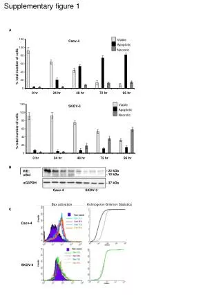
Mitochondrial targeting of TR3 regulates mitochondrial activity. (A) TR3 expression is required for cytochrome c release in response to apoptosis inducers.(B) Mitochondrial targeting of TR3 is associated with cytochrome c release.(C) TR3/ 1 blocks cytoplasmic localization of TR3. (D) TR3/ 1 inhibits MM11453-induced apoptosis. Quantification of apoptotic cells in 400TR3/ 1-transfected or nontransfected cells is shown in (E).
Mitchondrial response to apoptotic signals • Bax and Bak • Bcl-2 and other antiapoptotic members • VDAC and ANT
Bax and Bak Die during embryonic development most Bax and Bak double KO mice A persistence of 指间蹼 An imperforate vaginal canal An accumulation of excess cells in nervous and 造血系统 few Resistant to multiple apoptotic stimuli ( overexpression of tBid, Bim and Bad)
BH3-only proteins Bax and Bak (mit) Conformayional changes and oligomerization(Bax and Bak) ? Form a big pore Destabilize the mitchondrial outer membrane Apoptogenic proteins(cytochrome c, Smac, EndoG )pass through
Bcl-2 Two steps in the Bax apoptotic pathway
Bcl-2 and other antiapoptotic members BH3-only proteins X Bcl-2, Bcl-xL Bax and Bak Bcl-2, Bcl-xL oligomerization apoptosis
VDAC and ANT VDAC and ANT (outer and inner membranes of mit) Bax + Small metabolites and nucleotides across VDAC-Bax ANT-Bax - cytokine Export 肌酸磷酸 Form protein pore Change of VDAC permeability ATP/ADP exchage Cyto c release ? apoptosis
Loss of mitchondrial functions during apoptosis Apoptotic stimuli Suffer special damage (mit) Loss of mit function apoptosis
Release of cyto c Maintain membrane potential ATP synthesis Electron transfer Overexpression of Bcl-xL mit ADP和内膜肌 酸磷酸脂 浓度 ATP Loss of mitochondrial function BH3-only proteins Abolish the ability to buffer cacium, important mit function for cellular homeostasis tBid +cyto c in early stage
Cyto c release AIF, EndoG release ATP synthesis interruption - caspases activation Mit dysfunction + caspases activation 代谢变化(1)Mit matrix alkalinization; 2)谷胱肝肽-S 转移酶; 果糖1,6-二磷酸;脂肪酸结合蛋白;UPC-2;VDAC活性 apoptosis
perspectives • How are the signals from either developmental cues or damage signals transduced to and integratedin the mit? • Are the BH3-only proteins the major signal transducers? Or are they only part of a more complicated network of proteins?