Download
1 / 90
930 likes | 1.38k Views
Skull and Face. Boonsirm Withyachumnarnkul, M.D., Ph.D. Department of Anatomy, Faculty of Science Mahidol University. Bony Landmarks of Skull and Face. Vertex Superciliary arch Zygoma Mental symphysis Entrance to orbit Anterior nasal aperture. Three foramina vertically alligned.

E N D
Skull and Face Boonsirm Withyachumnarnkul, M.D., Ph.D. Department of Anatomy, Faculty of Science Mahidol University
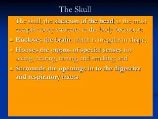
Bony Landmarks of Skull and Face • Vertex • Superciliary arch • Zygoma • Mental symphysis • Entrance to orbit • Anterior nasal aperture
Three foramina vertically alligned • Supraorbital foramen … for supraorbital nerve • Infraorbital foramen … for infraorbital nerve • Mental foramen ... for mental nerve
Muscles of Facial Expression • Develop from the 2nd branchial arch, thus all are supplied by CN VII. • Most are thin, originate from facial bones to insert on facial skin, except platysma, and are intermingled at their insertions.
Muscles of Facial Expression • Muscle of the forehead … frontalis, as part of the occipitofrontalis • Muscles of the mouth • Muscle of the eyelids • Muscle of the nose • Platysma
Muscles of the Mouth • Orbicularis oris • Zygomaticus major • Zygomaticus minor • Levator labii superioris • Levator labii superioris alaque nasi • Buccinator • Depressor anguli oris • Depressor labii inferioris • Mentalis • Risorius • Platysma
Muscle of the Eyelids • Orbicularis oculi • Orbital part • Palpebral part
Buccal Pad Fat • Between masseter and buccinator muscles • Brown fat … for heat generation, especially for children
Parotid Duct • One finger-breadth below zygomatic arch • Open into the mouth cavity (vestibule) at the level of the 2nd molar tooth (crown)
Facial Nerve • Comes out from stylomastoid foramen • Five branches • Temporal • Zygomatic • Buccal • Mandibular • Cervical
Facial Palcy • No wrinkle of forehead • Angle of mouth drops • Sagging lower eyelid • Other signs relating to malfunctions of structures innervated by facial nerve
Trigeminal Nerve • Sensory • Ophthalmic division • Maxillary division • Mandibular division • Motor .. To 1st branchial arch • Muscles of mastication • Messeter • Temporalis • Medial pterygoid • Lateral pterygoid
Arteries of the Face(rich, tortuous and highly anastomosed) • From external carotid artery • Facial artery • Superficial temporal artery • Transverse facial artery • From internal carotid artery • supraorbital artery • supratrochlear artery
Veins of the Face • Two important veins • Facial vein • Retromandibular vein • Facial veins have no valves • Connection of facial veins, pterygoid plexus and cavernous sinus
Lymph Drainage of the Face • Submental lymph nodes • Submandibular lymph nodes • Parotid lymph nodes
Scalp, Cranial Cavity and Venous Sinuses Boonsirm Withyachumnarnkul, M.D., Ph.D. Department of Anatomy, Faculty of Science Mahidol University Head2.ppt in C (Mahidol)
Scalp • Five layers of scalp • skin • dense subcutaneous tissue • epicranial aponeurosis • loose areolar connective tissue • periosteum
Scalp • Clinical relevance • infection spreading from loose areolar connective tissue, via emissary veins, to meninges-meningitis • hematoma
Skull Cap or Calvaria • Suture • coronal (frontoparietal) • anterior fontanelle • sagittal (interparietal) • lambdoid (occipitoparietal) • posterior fontanelle
Skull Cap or Calvaria • Three layers of skull cap • outer table • diploe • inner table
Cranial Fossae • Anterior cranial fossa • Middle cranial fossa • Posterior cranial fossa • Boundaries • lesser wing of sphenoid • superior border of petrous bone
Anterior Cranial Fossa • important landmarks • crista galli & cribriform plate of ethmoid • sella turcica • tuberculum sellae • hypophyseal fossa • dorsum sellae
Middle Cranial Fossa • important landmarks • foramina • superior orbital fissure • foramen rotundum • foramen ovale • foramne spinosum • groove for middle meningeal artery
Posterior Cranial Fossa • important landmarks • grooves for transverse & sigmoid sinuses • foramen magnum
Dura Mater • Outer and inner layers • position of the middle meningeal artery
Dura Mater • Falx cerebri • Falx cerebelli • Tentorial cerebelli and notch • Diaphragmatic sellae
Intradural Venous Sinuses • Superior and inferior sagittal sinuses • Straight sinus • Transverse and sigmoid sinuses
Intradural Venous Sinuses • Cavernous sinus • relationship among internal carotid artery, CN III, CN IV, CN V1 and CN VI • venous connections • clinical relevance • thrombophlebitis
Orbit and Eye Boonsirm Withyachumnarnkul, M.D., Ph.D. Department of Anatomy, Faculty of Science Mahidol University
Eye From the Outside • eyelids • palpebral fissure • plica semilunaris • caruncle • lacrimal puncta • cornea • sclera • conjunctiva • bulbar • palpebral • Sty • pterygium
Bony Parts of the Orbit • Entrance of the Orbit • frontal bone • zygomatic bone • maxillary bone • More Bones Inside • ethmoid bone • greater and lesser wing of sphenoid • lacrimal bone
Foramina of the Orbit • optic foramen (canal) • optic n. • ophthalmic a. • superior orbital fissure • all other nerves • superior ophthalmic vein • inferior orbital fissure • infraorbital n. & a. • inferior ophthalmic v. • infraorbital groove & foramen • zygomatic & infraorbital n. • supraorbital notch & foramen • supraorbital n.
Eyeball • Muscles • Extrinsic (Extra-ocular) • Intrinsic
Levator Palpebrae Superioris • Supplied by CN III • insert on the upper lid • if paralyzed ptosis
Superior Rectus Inferior Rectus Lateral Rectus Medial Rectus Superior Oblique Inferior Oblique Extra-Ocular Muscles
Actions of the Extra-Ocular Muscles • Around vertical axis • medial or adduction • lateral or abduction • Around horizontal axis • upward or elevation • downward or depression • Around antero-posterior axis • medial rotation • lateral rotation
Superior Rectus Make a 10-15 o with an AP axis adduct medial rotate elevate
Inferior Rectus depress adduct lateral rotate
Medial and Lateral Recti • Medial Rectus • adduction • Lateral Rectus • abduction
Superior Oblique • depress • abduction • medial rotate
Inferior Oblique • elevate • abduction • lateral rotate
Nerves of the Extra-Ocular Muscles • Oculomotor Nerve (CN III) • supplies all except lateral rectus and superior oblique • Trochlear Nerve (CN IV) • superior oblique • Abducens Nerve (CN VI) • lateral rectus
Oculomotor nerve (CN III) Superior Division • levator palpebrae superioris* • superior rectus Inferior Division • inferior rectus • inferior oblique • medial rectus *not an extra-ocular muscle
Functional Tests of the Extra-Ocular Muscle • Principle • align the muscle axis with the eyeball AP axis • contract the muscle • e.g., for Superior Rectus • abduct, first • then, elevate • Therefore, test for the superior rectus function is to abduct and elevate
Parts of the Eyeball • Three layers • sclera • choroid • retina • anterior chamber • posterior chamber • cornea • iris • ciliary muscle • suspensory ligament • lens • hyaloid canal • vitreous body • aqueous humor
Clinical Relevance • lenticular cataract • glaucoma • Schlemn’s canal • Myopia (near sightedness) • hyperopia (far sightedness) • presbiopia (old-aged sightedness)