Download

1 / 161
1.65k likes | 1.9k Views
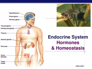
Nerves, Hormones, and Homeostasis. Heinemann.co.uk/hotlinks Express code: 4273P. The Nervous System Links Sensation to Response. Structure and Function of the Nervous System Neurons (Nerve Cells) – Basic Unit of the nervous

E N D
Nerves, Hormones, and Homeostasis • Heinemann.co.uk/hotlinks • Express code: 4273P
The Nervous System Links Sensation to Response Structure and Function of the Nervous System Neurons (Nerve Cells) – Basic Unit of the nervous system • Neurons are specialized to carry electrical signals
Nervous system has two subsystems: • Central Nervous System (CNS) • The body’s main information Processing center
Nervous system has two subsystems: • Peripheral Nervous System (PNS) • Outside the CNS • PNS delivers information to the CNS and carries messages from the CNS to other organs Nerve – Consist of one or more bundles of neuron fibers surrounded by connective tissue
Two Categories of Peripheral Nerve • Spinal Nerve – 31 pairs (left and right) emerge from the spinal cord. They are mixed nerves, some sensory and some motor • Cranial Nerves – 12 pairs of these emerge from an area of the brain known as the brainstem. • I.e. The optic nerve – that take visual info. from the retina to the brain.
Stimulation & Interpretation There are many types of nerve receptors • When touched: • That touch or pressure stimulates an action potential • This information reaches the spinal cord • Sensory neurons stretch from receptors to the spinal cord
Stimulation & Interpretation • Once the action potential reaches the spinal cord, it is routed in the CNS to the appropriate area for interpretation • Touch, pain, pressure, etc. Relay Neurons – neurons that carry impulses within the spinal cord and brain
Stimulation & Interpretation Response • Interpretation is made by the appropriate area of the brain • Reaction to a stimulus • Deciding to move: • The brain’s relay neurons pass the action to spinal nerve pairs • Action potential is now on a pathway of motor neurons (neurons taking an impulse to a muscle) • Spinal nerve contains both sensory and motor neurons
Stimulation & Interpretation Response Motor End Plate – the junction which a neuron sends a chemical to muscle tissue which results in a contraction • Action potential reaches the motor end plate causing the muscle contraction initiating a response Effector – muscle that contracts because of the message to contract
3 Main Functions of the PNS and the CNS working together: • Sensory Input Stimulus– (plural, stimuli) information received from the environment by a receptor and elicits a response • Information received by the PNS • Examples: colors, change in temperature, being touched Sensory Neurons – neurons that carry information about stimuli, to the CNS.
Main Functions of the PNS and the CNS Sensory Input Sensory Receptors – highly specialized cells that receive stimuli.
Main Functions of the PNS and the CNS • Integration • CNS interprets the information • Neurons are located entirely within the CNS Interneuron – neurons in the CNS, integrates sensory input
Main Functions of the PNS and the CNS • Motor Output Motor Neurons - neurons that carry signals away from CNS • Like moving an arm / Throwing a ball
Main Functions of the PNS and the CNS Reflex – a rapid, automatic (unconscious) response without thinking Reflex Arc – the nervous system pathway that regulates a reflex
Reflex : • Receptors detect / receive stimulus • Pain receptors receive the stimulus of excess heat, pressure, or chemicals produced by injured tissues • Sensory neurons in the PNS convey this information to the CNS (spinal cord) • The axon of the sensory neuron enters the spinal cord in the dorsal root and sends a chemical message across synapse to a relay neuron
Reflex • Relay neuron is located in the grey matter of the spinal cord • White matter occurs wherever conduction of impulses is the major event • Grey matter occurs wherever integration of impulses may occur • CNS (spinal cord) transmit signals to both motor neurons and interneuron • Relay neuron synapse with a motor neuron in the grey matter and transfers the impulse is transferred chemically across the synapse
Reflex • The motor neuron is located in the ventral root of the spinal cord • The motor neuron Is located in the ventral root of the spinal cord • It carries the impulse to an effector
Effects of Natural Selection • Animal behavior can change in response to their environment • The behaviors can be so extreme that a new species is formed • Variations in behavior can occur in population in the same way variations can occur in appearance • Variations in behavior can be selected by the environment
Effects of Natural Selection • Genetically programmed behavior can have variations that can work better than another in a changing environment • Causing that organism to survive and reproduce • Read page 339 - 340 about European Blackcaps & Sockeye Salmon
Neurons Conduct Nerve Impulses Structure of a Neuron Cell Body – Contains neuron’s nucleus and most organelles Dendrites – Receive signals and carry them towards the neuron’s cell body Axon – Carries electrical impulses away from the cell body and toward other cells. • Some axons can be very long, like from spinal cord to the toes.
Structure of a Neuron Myelin Sheath – Axons are insulated by a thick coat of material; they resemble a chain of oblong beads. Nodes – Areas between the myelin sheath (beads) that are un-insulated. • Signals will jump from node to node
A Neuron at Rest To better understand how a neuron works, first you need to understand a neuron at rest
Resting Potential Non-myelinatedNeurone – an axom that does not contain a myelinated sheath • Myelinated sheath speeds up the action potential Resting Potential – The state of being where a neuron is ready to send an action potential • This area of the neuron is said to be polarized
Resting Potential • Key is the plasma membrane: • Separates ions inside and outside the cell (ions electrically charged atoms / molecules) • Because opposite charges attract, separating them is a form of potential energy
Resting Potential Voltage – the “pressure” created when holding opposite charges apart. • Measured in units called volts • A resting neuron has voltage of -70 mV • The (-) indicates that the inside of the nerve cell is negative in charge compared to the outside.
Resting Potential Resting Potential – the voltage across the plasma membrane of a resting neuron. • Potential = potential energy
Ion Channels • The ions we are talking about in this Resting Potential are Na⁺ (sodium ions) outside • K⁺ (potassium ions) are more concentrated inside • Specific proteins in the membrane act like channels for each ion • So….. Na⁺ goes in cell; K⁺ goes out • There are more K⁺ channels than Na⁺ • So K⁺ goes out faster than Na⁺ goes in • This “unbalance” causes the outside of cell to be more (+) than inside • In addition there are negatively charged ions inside the cell
Ion Pumps Sodium Potassium Pump • A protein on the membrane (of neuron) that pumps ions across the membrane • Requires ATP • The resting potential creates a resting neuron that is ready to fire / transmit a nerve signal
How a Nerve Signal Travels • All cells, not just neurons, have voltages across their membrane • Only muscles and nerve cells can use this energy
Triggering the Nerve Signal Depolarization – if a neuron is stimulated, the voltage across the membrane changes at the point of stimulation. • Charge difference decreases across the membrane decreases. • Stimulation causes Na⁺ diffuses into the cell.
Triggering the Nerve Signal • Normally there is more Na⁺ outside the cell membrane • inflow of Na⁺ ions (⁺ charge) depolarizes the membrane
Triggering the Nerve Signal Threshold – the point when stimulus is strong enough to depolarize the membrane to a certain level. • Usually – 50mV • Additional Na⁺ channels open • Then there is a “rush” of Na⁺ into the cell • Causes greater depolarization
Triggering the Nerve Signal Action Potential – This is a stronger depolarization and the start of the nerve signal.
Transmitting the Nerve Signal • The 1st action Potential; causes Na⁺ gates to open nearby, causing more action potential; and so on…. • Like tipping over the first domino in a chain • Self – propagating part of action potential; once you start an impulse at the dendrite end, that action potential will self propagate itself to the far axon end of the cell
Transmitting the Nerve Signal • After a nerve signal passes a region of the neuron the resting potential is restored • The return to resting potential is caused by opening K⁺ gates • During action potential; concentration differences of Na⁺ and K⁺ ions Repolarization – the active transport is required to pump the two ions to their resting potential positions • This happens very quickly
The Speed of Transmission • 5 meters per second – along neurons membrane • Faster Even: because of myelin sheath
The Speed of Transmission • Action potential “jumps” from nodes to nodes • Makes nerve impulse faster • Neurons with myelin sheath transmits signals at 150 meters per second • From Spinal Cord to Toes in 7 milliseconds
Measuring Stimulus Strength • Action Potential = nerve signals; are all or none events • All nerve signals are equal • A stimulus becomes more intense when the frequency and the number of action potential increase
Measuring Stimulus Strength • The brain “reads” the number of action potentials to determine signal strength Refractory Period – the time it takes for any one neuron to send an action potential so it can send another
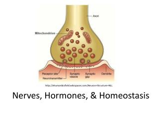
Crossing Synapses Synapse – the space between nerve cells, transmitting information / communicating • The first neuron is called presynaptic neuron • The second neuron is called postsynaptic neuron • Synapses can be chemical or electrical
Crossing Synapses • There are different patterns in presynaptic and postsynaptic communications • Can be one to one communication • Can be one presynaptic to many postsynaptic • Can be many presynaptic to one postsynaptic Action Potential
Crossing Synapses • A sensory pathway is unidirectional only because they are lined up so that the terminal end of the axon of the first neuron adjoins the dendrites of the next neuron.
Crossing Synapses Electrical Synapse – the action potential at the end of axon directly causes an electrical change in receiving cell • Common in the heart and digestive organs • Steady impulses are needed • Rhythmic muscle contractions
Crossing Synapses Chemical Synapse – nerve impulse must be transmitted across a tiny space.
Crossing Synapses Synaptic Cleft – space between knoblike tips and dendrite. • In chemical synapse the electrical signal is converted to a chemical signal. Terminal Buttons – located at the at the far end of axons are swollen areas • Area contains vesicles containing neurotransmitters
Crossing Synapses Neurotransmitters – Nitrogen-containing organic compounds, used to transmit signals across the synaptic cleft. When action potential reaches the knob: • Calcium ions diffuse into the terminal buttons • Vesicles are released into synaptic cleft by fusing with plasma membrane • NT diffuses across the synaptic gap