Download
1 / 17
2.1k likes | 7.57k Views
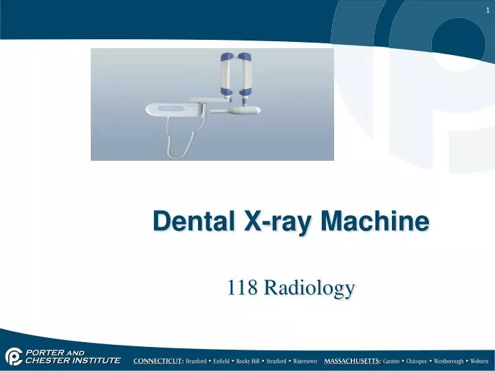
Dental X-ray Machine. 118 Radiology. Component Parts. The Dental x-ray machine consists of 3 visible component parts: The Control Panel The Extension Arm The Tubehead. Newer Design. Control Panels. Older style. The Control Panel. The Control Panel contains:

E N D
Dental X-ray Machine 118 Radiology
Component Parts • The Dental x-ray machine consists of 3 visible component parts: • The Control Panel • The Extension Arm • The Tubehead
Newer Design Control Panels Older style
The Control Panel • The Control Panel contains: • On-off switch and an indicator light • The exposure button and an indicator light • Control devices to regulate the x-ray beam • Time setting • kVp setting • mA setting
Miliamperage and Kilovoltage • Miliamperage = MA • controls number of electrons produced at tungsten filament quantity • 7-15 are average dental x-ray unit mA numbers • Kilovoltage = kV • controls penetrating power of electrons and x-ray quality • how fast they travel from cathode to anode • creates contrast on resulting film image • usually 70 or 90 kVpkilovoltage power
Extension Arm Hollow, holds electrical wires connecting tubehead with control panel Used to position tubehead Tubehead attached to arm by yoke, which can be moved 360 degrees horizontally Folds up when not in use
Tubehead Houses the x-ray tube Made of metal; lined with lead to prevent radiation leakage Filled with oil to absorb heat which is generated during x-ray production
X-ray Tube Made of glass 6” long x 1 ½” diameter Air is removed to create vacuum to allow electrons to flow freely between cathode and anode
Cathode Tungsten filament gets hot when electricity is sent to tube Creates electron cloud Focusing cup: Keeps electrons suspended at cathode When exposure button is pushed, electrons shoot across tube to anode Negative end of tube
Anode Postive end of tube • Tunsten target • Electrons strike target and product x-rays • 99% get absorbed by oil • 1% exits the tube at the area towards the patient • Center of this beam of x-rays is called the Central Ray
PID • Position indicating device • Lead lined • Aims x-ray beam at film in pt mouth OR at extra oral cassette • Open end placed against pt face • 8,12,16 inches long
Filter • Aluminum disc at port where the PID is connected to the Tubehead • Removes (absorbs) low-energy, long wavelength x-rays • Only allows high energy, short wave length x-rays to pas through • Both kinds of x-rays are absorbed by the pt’s tissues; only the short wave length , high energy x-rays create image on film
Collimator • Metal disc (lead) with small opening in center to control size and shape of x-ray beam • Further reduces pt exposure • 2 ¾” diameter; round shape and size • Also available in rectangular shape which exactly fits #2 film size exposes over ½ less tissue with rectangular collimator