Download

1 / 27
710 likes | 3.34k Views
ANATOMY OF THE FEMALE REPRODUCTIVE SYSTEM. Prof . Ahmed Fathalla Ibrahim Professor of Anatomy College of Medicine King Saud University E-mail: ahmedfathala@hotmail.com. OBJECTIVES. At the end of the lecture, students should: List the organs of female reproductive system.

E N D
ANATOMY OF THE FEMALE REPRODUCTIVE SYSTEM Prof. Ahmed FathallaIbrahim Professor of Anatomy College of Medicine King Saud University E-mail: ahmedfathala@hotmail.com
OBJECTIVES At the end of the lecture, students should: • List the organs of female reproductive system. • Describe the pelvic peritoneum in female. • Describe the position and relations of the ovaries. • List the parts of the uterine tube. • Describe the anatomy of uterus regarding: subdivisions, cavity, relations, ligaments & main support. • Describe the anatomy of vagina regarding: structure, extent, length & relations. • Describe the supply (arteries, veins, lymph, nerves) of female reproductive system.
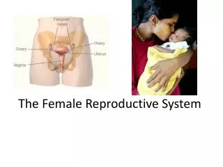
FEMALE REPRODUCTIVE SYSTEM UTERINE TUBE OVARY UTERUS V A G I N A POSTERIOR VIEW
PELVIC PERITONEUM IN FEMALE Rectouterine (Douglas) pouch: Reflection of peritoneum from rectum to upper part of posterior surface of vagina Uterovesical (vesicouterine) pouch: Reflection of peritoneum from uterus to upper surface of urinary bladder Broad ligament of uterus: Extension of peritoneum from lateral wall of uterus to lateral wall of pelvis, encloses the uterine tubes BROAD LIGAMENT
THE OVARIES It is an almond-shaped organ. It is attached to the back of the broad ligament by a peritoneal fold (mesovarium) Its medial end is attached to uterus by ligament of ovary. Its lateral end is related to the fimbriae of the uterine tube. BROAD LIGAMENT Female pelvic organs (Posterior view)
THE UTERINE (FALLOPIAN) TUBES 2 3 It is 10 cm long. It is enclosed in the broad ligament of uterus. It is divided into: 1) Intramural part: opening into the uterine wall 2)Isthmus: narrowest part 3)Ampulla: widest part (site of fertilization) 4)Infundibulum: funnel-shaped end, has finger-like processes (fimbriae), related to ovary 1 4 X
FUNCTION OF: OVARIES: PRIMARY SEX ORGANS IN FEMALE Production of female germ cells Secretion of female sex hormones UTERINE TUBES: Site of fertilization Transport of fertilized ovum into the uterus
2’ THE UTERUS 3’ A hollow, pear-shaped muscular organ Divided into: Fundus: no cavity Body: cavity is triangular Cervix: cavity is fusiform, divided into: *Supravaginal part *Vaginal part 1’ Fundus Body
THE UTERUS FUNDUS: The part of uterus above the level of uterine tubes BODY: The part of uterus from the level of uterine tube to the level of the isthmus of uterus CERVIX:The part of the uterus below the level of the isthmus of the uterus
THE CERVICAL CANAL INTERNAL OS: opening between cavity of body of uterus & cavity of cervix (cervical canal) EXTERNAL OS: opening between cervical canal & cavity of vagina In a nulliparous woman: external os appears circular. In a multiparous woman: external os appears as a transverse slit with an anterior & a posterior lip. Multiparous Nulliparous
RELATIONS OF UTERUS FUNDUS + BODY + SUPRAVAGINAL PART OF CERVIX: Anterior: superior surface of urinary bladder Posterior: sigmoid colon Lateral: uterine artery VAGINAL PART OF CERVIX: surrounded by vaginal fornices Anterior: anterior fornix of vagina Posterior: posterior fornix of vagina Lateral: lateral fornices of vagina
POSITIONS OF UTERUS ANTEFLEXED UTERUS ANTEVERTED UTERUS Long axis of body of uterus is bent forward on long axis of cervix Long axis of whole uterus is bent forward on long axis of vagina
POSITIONS OF UTERUS RETROVERTED UTERUS RETROFLEXED UTERUS Fundus & body of uterus are bent backward on the vagina and lie in rectouterine pouch Long axis of body of uterus is bent backward on long axis of cervix
USUAL POSITION OF UTERUS ANTEVERTED ANTEFLEXED UTERUS
LIGAMENTS OF UTERUS 1)Ligaments at junction between fundus & body of uterus (At the level of uterine tube) 2 Extends through inguinal canal to labium majus 1
LIGAMENTS OF UTERUS 2)Ligaments of cervix Extend from cervixto: anterior (pubocervical), lateral (transverse cervical or cardinal) posterior (uterosacral or sacrocervical) pelvic walls
LEVATOR ANI MUSCLES • FORM THE PELVIC FLOOR: separate pelvis from perineum • FORM PELVIC DIAPHRAGM: traversed by urethra, vagina & rectum • SUPPORT PELVIC ORGANS Urethra Levatorani Vagina Urethra Rectum Rectum Vagina
SUPPORT OF UTERUS • Ligaments of cervix (especially transverse cervical) • Round ligament of uterus (maintains antevertedanteflexed position) • Levatorani muscles UTERINE PROLAPSE • Downward dispalcement of uterus due to damage of: • Ligaments of uterus • Levatorani muscles
VAGINA • STRUCTURE: Fibro-muscular tube • EXTENT: From external os, along pelvis & perineum, to open in the vulva (female external genitalia), behind urethral opening • LENGTH: Its anterior wall (7.5 cm) is shorter than its posterior wall (9 cm) • FUNCTION: 1) Copulatory organ & 2) Birth canal
RELATIONS OF VAGINA • ANTERIOR: Urinary bladder (in pelvis) & urethra (in perineum) • POSTERIOR: Rectum (in pelvis) & anal canal (in perineum) • LATERAL:ureters(in pelvis)
ARTERIAL SUPPLY From abdominal aorta From internal iliac artery (Artery of pelvis)
QUESTION 1 • Which one of the following structures is related (or attached) to the lateral end of the ovary? • Fimbriae of uterine tube • Ampulla of uterine tube • Ligament of ovary • Round ligament of uterus
QUESTION 2 • Which one of the following structures is anterior to the uterus? • Urinary bladder • Ureter • Sigmoid colon • Ovary