Download
1 / 52
530 likes | 772 Views
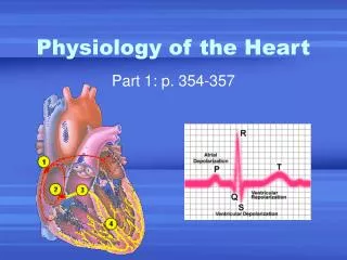
AP 151 Physiology of the Heart. Functions of the Heart. Generating blood pressure Routing blood: separates pulmonary and systemic circulations Ensuring one-way blood flow: valves Regulating blood supply Changes in contraction rate and force match blood delivery to changing metabolic needs.

E N D
AP 151 Physiology of the Heart
Functions of the Heart • Generating blood pressure • Routing blood:separates pulmonary and systemic circulations • Ensuring one-way blood flow: valves • Regulating blood supply • Changes in contraction rate and force match blood delivery to changing metabolic needs
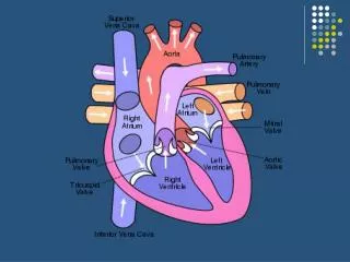
The cardiovascular system is divided into two circuits • Pulmonary circuit • blood to and from the lungs • Systemic circuit • blood to and from the rest of the body • Vessels carry the blood through the circuits • Arteries carry blood away from the heart • Veins carry blood to the heart • Capillaries permit exchange
Cardiac Muscle • Elongated, branching cells containing 1-2 centrally located nuclei • Contains actin and myosin myofilaments • Intercalated disks: specialized cell-cell contacts. • Cell membranes interdigitate • Desmosomes hold cells together • Gap junctions allow action potentials to move from one cell to the next. • Electrically, cardiac muscle of the atria and of the ventricles behaves as single unit • Mitochondria comprise 30% of volume of the cell vs. 2% in skeletal
Heart chambers and valves • Structural Differences in heart chambers • The left side of the heart is more muscular than the right side • Functions of valves • AV valves prevent backflow of blood from the ventricles to the atria • Semilunar valves prevent backflow into the ventricles from the pulmonary trunk and aorta
Cardiac Muscle Contraction • Heart muscle: • Is stimulated by nerves and is self-excitable (automaticity) • Contracts as a unit; no motor units • Has a long (250 ms) absolute refractory period • Cardiac muscle contraction is similar to skeletal muscle contraction, i.e., sliding-filaments
Differences Between Skeletal and Cardiac Muscle Physiology • Action Potential • Cardiac: Action potentials conducted from cell to cell. • Skeletal, action potential conducted along length of single fiber • Rate of Action Potential Propagation • Slow in cardiac muscle because of gap junctions and small diameter of fibers. • Faster in skeletal muscle due to larger diameter fibers. • Calcium release • Calcium-induced calcium release (CICR) in cardiac • Movement of extracellular Ca2+ through plasma membrane and T tubules into sarcoplasm stimulates release of Ca2+ from sarcoplasmic reticulum • Action potential in T-tubule stimulates Ca++ release from sarco-plasmic reticulum
The Action Potential in Skeletal and Cardiac Muscle Figure 20.15
Electrical Properties of Myocardial Fibers 1. Rising phase of action potential Due to opening of fast Na+ channels 2. Plateau phase Closure of sodium channels Opening of calcium channels Slight increase in K+ permeability Prevents summation and thus tetanus of cardiac muscle 3. Repolarization phase Calcium channels closed Increased K+ permeability
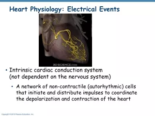
Conduction System of the Heart • SA node: sinoatrial node. The pacemaker. • Specialized cardiac muscle cells. • Generate spontaneous action potentials (autorhythmic tissue). • Action potentials pass to atrial muscle cells and to the AV node • AV node: atrioventricular node. • Action potentials conducted more slowly here than in any other part of system. • Ensures ventricles receive signal to contract after atria have contracted • AV bundle: passes through hole in cardiac skeleton to reach interventricular septum • Right and left bundle branches: extend beneath endocardium to apices of right and left ventricles • Purkinje fibers: • Large diameter cardiac muscle cells with few myofibrils. • Many gap junctions. • Conduct action potential to ventricular muscle cells (myocardium)
Heart Physiology: Intrinsic Conduction System • Autorhythmic cells: • Initiate action potentials • Have unstable resting potentials called pacemaker potentials • Use calcium influx (rather than sodium) for rising phase of the action potential
Depolarization of SA Node • SA node - no stable resting membrane potential • Pacemaker potential • gradual depolarization from -60 mV, slow influx of Na+ • Action potential • occurs at threshold of -40 mV • depolarizing phase to 0 mV • fast Ca2+ channels open, (Ca2+ in) • repolarizing phase • K+ channels open, (K+ out) • at-60 mV K+ channels close, pacemaker potential starts over • Each depolarization creates one heartbeat • SA node at rest fires at 0.8 sec, about 75 bpm
Heart Physiology: Sequence of Excitation • Sinoatrial (SA) node generates impulses about 75 times/minute • Atrioventricular (AV) node delays the impulse approximately 0.1 second • Impulse passes from atria to ventricles via the atrioventricular bundle (bundle of His) to the Purkinje fibers and finally to the myocardial fibers
Electrocardiogram • Record of electrical events in the myocardium that can be correlated with mechanical events • P wave: depolarization of atrial myocardium. • Signals onset of atrial contraction • QRS complex: ventricular depolarization • Signals onset of ventricular contraction.. • T wave: repolarization of ventricles • PR interval or PQ interval: 0.16 sec • Extends from start of atrial depolarization to start of ventricular depolarization (QRS complex) contract and begin to relax • Can indicate damage to conducting pathway or AV node if greater than 0.20 sec (200 msec) • Q-T interval: time required for ventricles to undergo a single cycle of depolarization and repolarization • Can be lengthened by electrolyte disturbances, conduction problems, coronary ischemia, myocardial damage
ECGs, Abnormal Extrasystole : note inverted QRS complex, misshapen QRS and T and absence of a P wave preceding this contraction.
ECGs, Abnormal Arrhythmia: conduction failure at AV node No pumping action occurs
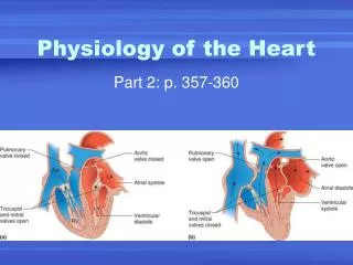
The Cardiac Cycle • Cardiac cycle refers to all events associated with blood flow through the heart from the start of one heartbeat to the beginning of the next • During a cardiac cycle • Each heart chamber goes through systole and diastole • Correct pressure relationships are dependent on careful timing of contractions
Phases of the Cardiac Cycle • Atrial diastole and systole - • Blood flows into and passively out of atria (80% of total) • AV valves open • Atrial systole pumps only about 20% of blood into ventricles • Ventricular filling: mid-to-late diastole • Heart blood pressure is low as blood enters atria and flows into ventricles • 80% of blood enters ventricles passively • AV valves are open, then atrial systole occurs • Atrial systole pumps remaining 20% of blood into ventricles
Phases of the Cardiac Cycle • Ventricular systole • Atria relax • Rising ventricular pressure results in closing of AV valves (1st heart sound - ‘lubb’) • Isovolumetric contraction phase • Ventricles are contracting but no blood is leaving • Ventricular pressure not great enough to open semilunar valves • Ventricular ejection phase opens semilunar valves • Ventricular pressure now greater than pressure in arteries (aorta and pulmonary trunk)
Phases of the Cardiac Cycle • Ventricular diastole • Ventricles relax • Backflow of blood in aorta and pulmonary trunk closes semilunar valves (2nd hear sound - “dubb • Dicrotic notch – brief rise in aortic pressure caused by backflow of blood rebounding off semilunar valves • Blood once again flowing into relaxed atria and passively into ventricles
Cardiac Output (CO) and Cardiac Reserve • CO is the amount of blood pumped by each ventricle in one minute • CO is the product of heart rate (HR) and stroke volume (SV) CO = HR x SV (ml/min) = (beats/min) x ml/beat • HR is the number of heart beats per minute • SV is the amount of blood pumped out by a ventricle with each beat • Cardiac reserve is the difference between resting and maximal CO
A Simple Model of Stroke Volume Figure 20.19a-d
Cardiac Output: An Example • CO (ml/min) = HR (75 beats/min) x SV (70 ml/beat) • CO = 5250 ml/min (5.25 L/min) • If HR increases to 150 b/min and SV increases to 120 ml/beat, then • CO = 150 b/min x 120 ml/beat • CO = 18,000 ml/min or 18 L/min (WOW is right!!)
Factors Affecting Cardiac Output Figure 20.20
Heart Rate • Pulse = surge of pressure in artery • infants have HR of 120 bpm or more • young adult females avg. 72 - 80 bpm • young adult males avg. 64 to 72 bpm • HR rises again in the elderly • Tachycardia: resting adult HR above 100 • stress, anxiety, drugs, heart disease or body temp. • Bradycardia: resting adult HR < 60 • in sleep and endurance trained athletes
Regulation of Heart Rate • Positive chronotropic factors increase heart rate • Chrono - time • Negative chronotropic factors decrease heart rate
Extrinsic Innervation of the Heart • Vital centers of medulla 1. Cardiac Center • Cardioaccelerator center • Activates sympathetic neurons that increase HR • Cardioinhibitory center • Activates parasympathetic neurons that decrease HR • Cardiac center receives input from higher centers (hypotha-lamus), monitoring blood pressure and dissolved gas concentrations
Regulation of the Heart • Neural regulation • Parasympathetic stimulation - a negative chronotropic factor • Supplied by vagus nerve, decreases heart rate, acetylcholine is secreted and hyperpolarizes the heart • Sympathetic stimulation - a positive chronotropic factor • Supplied by cardiac nerves. • Innervate the SA and AV nodes, and the atrial and ventricular myocardium. • Increases heart rate and force of contraction. • Epinephrine and norepinephrine released. • Increased heart beat causes increased cardiac output. Increased force of contraction causes a lower end-systolic volume; heart empties to a greater extent. Limitations: heart has to have time to fill. • Hormonal regulation • Epinephrine and norepinephrine from the adrenal medulla. • Occurs in response to increased physical activity, emotional excitement, stress
Basic heart rate established by pacemaker cells • SA node establishes baseline (sinus rhythmn) • Modified by ANS • If all ANS nerves to heart are cut, heart rate jumps to about 100 b/min • What does this tell you about which part of the ANS is most dominant during normal period?
Chemical Regulation of the Heart • The hormones epinephrine and thyroxine increase heart rate • Intra- and extracellular ion concentrations must be maintained for normal heart function
Regulation of Stroke Volume • SV: volume of blood pumped by a ventricle per beat SV= end diastolic volume (EDV) minus end systolic volume (ESV); SV = EDV - ESV • EDV = end diastolic volume • amount of blood in a ventricle at end of diastole • ESV = end systolic volume • amount of blood remaining in a ventricle after contraction • Ejection Fraction - % of EDV that is pumped by the ventricle; important clinical parameter • Ejection fraction should be about 55-60% or higher
Factors Affecting Stroke Volume • EDV - affected by • Venous return - vol. of blood returning to heart • Preload – amount ventricles are stretched by blood (=EDV) • ESV - affected by • Contractility – myocardial contractile force due to factors other than EDV • Afterload – back pressure exerted by blood in the large arteries leaving the heart
Frank-Starling Law of the Heart • Preload, or degree of stretch, of cardiac muscle cells before they contract is the critical factor controlling stroke volume; EDV leads to stretch of myocard. • preload stretch of muscle force of contraction SV • Unlike skeletal fibers, cardiac fibers contract MORE FORCEFULLY when stretched thus ejecting MORE BLOOD (SV) • If SV is increased, then ESV is decreased!! • Slow heartbeat and exercise increase venous return (VR) to the heart, increasing SV • VR changes in response to blood volume, skeletal muscle activity, alterations in cardiac output • VR EDV and in VR in EDV • Any in EDV in SV • Blood loss and extremely rapid heartbeat decrease SV
Extrinsic Factors Influencing Stroke Volume • Contractility is the increase in contractile strength, independent of stretch and EDV • Referred to as extrinsic since the influencing factor is from some external source • Increase in contractility comes from: • Increased sympathetic stimuli • Certain hormones • Ca2+ and some drugs • Agents/factors that decrease contractility include: • Acidosis • Increased extracellular K+ • Calcium channel blockers
Effects of Autonomic Activity on Contractility • Sympathetic stimulation • Release norepinephrine from symp. postganglionic fiber • Also, EP and NE from adrenal medulla • Have positive ionotropic effect • Ventricles contract more forcefully, increasing SV, increasing ejection fraction and decreasing ESV • Parasympathetic stimulation via Vagus Nerve -CNX • Releases ACh • Has a negative inotropic effect • Hyperpolarization and inhibition • Force of contractions is reduced, ejection fraction decreased
Contractility and Norepinephrine • Sympathetic stimulation releases norepinephrine and initiates a cyclic AMP 2nd-messenger system Figure 18.22
Preload and Afterload Figure 18.21
Effects of Hormones on Contractility • Epi, NE, and Thyroxine all have positive ionotropic effects and thus contractility • Digitalis elevates intracellular Ca++ concentrations by interfering with its removal from sarcoplasm of cardiac cells • Beta-blockers (propanolol, timolol) block beta-receptors and prevent sympathetic stimulation of heart (neg. chronotropic effect)
Exercise and Cardiac Output • Proprioceptors • HR at beginning of exercise due to signals from joints, muscles • Venous return • muscular activity venous return causes SV • HR and SV cause CO • Exercise produces ventricular hypertrophy • SV allows heart to beat more slowly at rest • cardiac reserve