Download
1 / 118
1.18k likes | 1.24k Views
This module covers reproductive anatomy, fetal development, pregnancy changes, terminology, and assessment. Learn about pelvic structure, reproductive cycles, and conception to fetal development stages. Understand the physiological, psychological, and nutritional aspects of pregnancy.

E N D
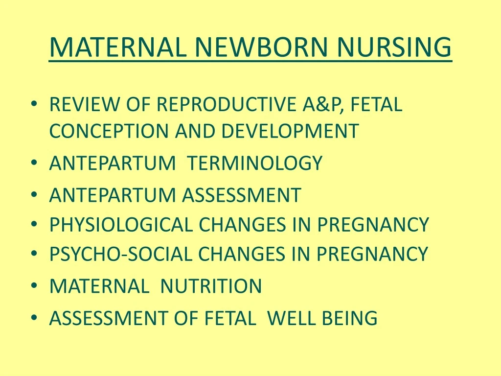
MATERNAL NEWBORN NURSING • REVIEW OF REPRODUCTIVE A&P, FETAL CONCEPTION AND DEVELOPMENT • ANTEPARTUM TERMINOLOGY • ANTEPARTUM ASSESSMENT • PHYSIOLOGICAL CHANGES IN PREGNANCY • PSYCHO-SOCIAL CHANGES IN PREGNANCY • MATERNAL NUTRITION • ASSESSMENT OF FETAL WELL BEING
MODULE 1 PART 1 REVIEW OF REPRODUCTIVE ANATOMY AND PHYSIOLOGY
REVIEW • REPRODUCTIVE A&P, FETAL CONCEPTION & DEVELOPMENT THIS WILL NOT BE COVERED IN THIS LECTURE—BE PREPARED TO ANSWER REVIEW QUESTIONS IN CLASS THE QUIZ IN CLASS 1 WILL FOCUS ON CHANGES IN PREGNANCY AND TERMINOLOGY
Review of Reproductive A&P • External Genitals • Internal Reproductive Organs • Vagina • Uterus • Uterine corpus • Cervix • Uterine ligaments • Fallopian Tubes • Ovaries
REVIEW OF REPRODUCTIVE A&P • UTERINE LIGAMENTS • ROUND LIGAMENTS • OVARIAN LIGAMENTS • CARDINAL LIGAMENTS • INFUNDIBULOPELVIC LIGAMENT • UTEROSACRAL LIGAMENT
Figure 2–3b Blood supply to vagina, ovaries, uterus, and fallopian tube.
Figure 2–3a Blood supply to internal reproductive organs. Pelvic blood supply.
PELVIC STRUCTURE • Innominate bones • ILIUM • ILIAC CREST • ISCHIUM • ISCHIAL TUBEROSITY • ISCHIAL SPINES • PUBIS • SYMPHYSIS PUBIS • Sacrum • SACRAL PROMOTORY, SACROILIAC JOINTS • Coccyx
REVIEW OF REPRODUCTIVE A&P • PELVIC DIVISION • TRUE PELVIS • INLET • PELVIC CAVITY • OUTLET
Figure 2–10a Female pelvis. False pelvis is shallow cavity above the inlet; true pelvis is deeper portion of cavity below the inlet.
Figure 2–11 Pelvic planes: coronal section and diameters of the bony pelvis.
REVIEW OF REPRODUCTIVE A&P • PELVIC DIAPHRAGM • LEVATOR ANI • COCCYGEAL MUSCLES • DEEP FASCIA • PELVIC FLOOR MUSCLES • LEVATOR ANI • ILLIOCOCCYGEUS • PUBOCOCCYGEUS, COCCYGEUS • PUBORECTALIS, PUBORECTALIS • PUBOVAGINALIS
Figure 2–9 Muscles of the pelvic floor. (The puborectalis, pubovaginalis, and coccygeal muscles cannot be seen from this view.)
REVIEW OF REPRODUCTIVE A&P • BREASTS • NIPPLE • AREOLA • TUBERCLES OF MONTGOMERY • LACTIFEROUS DUCTS • ADIPOSE, GLANDULAR, FIBROUS TISSUE • COOPER’S LIGAMENTS
Figure 2–12 Anatomy of the breast: sagittal view of left breast.
MATURATION OF OVARIAN FOLLICLE • OVULATION • CORPUS LUTEUM • NEUROHUMORAL RESPONSE • HYPOTHALMUS RELEASES GONADATROPIN-RELEASING HORMONE TO PITUITARY FROM RESPONES FROM CNS • ANTERIOR PITUITARY THEN SECRETES FSH AND LH
FEMALE REPRODUCTIVE CYCLE • OVARIAN CYCLE • FOLLICULAR PHASE • LUTEAL PHASE • FEMALE HORMONES • ESTROGEN • PROGESTERONE • PROSTAGLANDINS • UTERINE CYCLE (MENSTRUAL)
Figure 2–13 Female reproductive cycle: interrelationships of hormones with the four phases of the uterine cycle and the two phases of the ovarian cycle in an ideal 28-day cycle.
Figure 2–14 Various stages of development of the ovarian follicles.
REVIEW OF CONCEPTION AND FETAL DEVELOPMENT • CELLULAR DIVISION • MITOSIS • MEIOSIS • OOGENESIS • SPERMATOGENESIS • PRE-FERTILIZATION • CAPACIATION • ACROSOMAL REACTION • FERTILIZATION
PREEMBRYONIC STAGE • CELLULAR MULTIPLICATION • CLEAVAGE • MORULA • BLASTOCYST • TROPHOBLAST • IMPLANTATION • CHANGES IN ENDOMETRIUM • DECIDUA CAPSULARIS • DECIDUA BASALIS • DICIDUA VERA
Figure 3–2a Sperm penetration of an ovum. The sequential steps of oocyte penetration by a sperm are depicted moving from top to bottom. Source: Scanning electron micrograph from Nilsson, L. (1990). A child is born. New York: Dell Publishing.
Figure 3–1b Each spermatogonium produces four haploid spermatozoa.
Figure 3–5 Formation of primary germ layers. A, Implantation of a 71⁄2-day blastocyst in which the cells of the embryonic disc are separated from the amnion by a fluid-filled space. The erosion of the endometrium by the syncytiotrophoblast is ongoing. B, Implantation is completed by day 9, and extraembryonic mesoderm is beginning to form a discrete layer beneath the cytotrophoblast. C, By day 16 the embryo shows all three germ layers, a yolk sac, and an allantois (an outpouching of the yolk sac that forms the structural basis of the body stalk, or umbilical cord). The cytotrophoblast and associated mesoderm have become the chorion, and chorionic villi are developing. Source: Adapted from Marieb, E. N. (1998).
Figure 3–4 During ovulation, the ovum leaves the ovary and enters the fallopian tube. Fertilization generally occurs in the outer third of the fallopian tube. Subsequent changes in the fertilized ovum from conception to implantation are depicted.
CELLULAR DIFFERENTIATION • THREE PRIMARY GERM LAYERS • ECTODERM • MESODERM • ENDODERM • EMBRYONIC MEMBRANES • AMNION • CHORION • AMNIOTIC SAC
REVIEW FETAL DEVELOPMENT • AMNIOTIC FLUID • UMBILICAL CORD • PLACENTA
REVIEW OF FETAL DEVELOPMENT • EMBRYONIC AND FETAL DEVELOPMENT • EMBRYONIC STAGE—DAY 15 T0 8TH WEEK • FETAL STAGE—8TH WEEK TO BIRTH
Figure 3–10 Vascular arrangement of the placenta. Arrows indicate the direction of blood flow. Maternal blood flows through the uterine arteries to the intervillous spaces of the placenta and returns through the uterine veins to maternal circulation. Fetal blood flows through the umbilical arteries into the villous capillaries of the placenta and returns through the umbilical vein to the fetal circulation.
Figure 3–7 Early development of primary embryonic membranes. At 41⁄2 weeks, the decidua capsularis (placental portion enclosing the embryo on the uterine surface) and decidua basalis (placental portion encompassing the elaborate chorionic villi and maternal endometrium) are well formed. The chorionic villi lie in blood-filled intervillous spaces within the endometrium. The amnion and yolk sac are well developed. Source: Adapted from Marieb, E. N. (1998).
Figure 3–6 Endoderm differentiates to form the epithelial lining of the digestive and respiratory tracts and associated glands. Source: Adapted from Marieb, E. N. (1998).
Figure 3–12 The actual size of a human conceptus from fertilization to the early fetal stage. The embryonic stage begins in the third week after fertilization; the fetal stage begins in the ninth week. Source: Adapted from Marieb, E. N. (1998).
REVIEW QUESTIONS • WHAT IS THE SIGNIFICANCE OF THE ENDOMETRIAL (MUCOSAL) LAYER OF THE UTERUS? • THE UTERUS IS MADE UP OF WHAT TYPE OF MUSCLE? • ESTROGEN IS SECRETED BY THE_______? • PROGESTERONE IS SECRETED BY THE_______? • WHAT IS THE FUNCTION OF LSH AND FH? • DESCRIBE MEIOSIS.
REVIEW QUESTIONS • WHERE DOES FERTILIZATION OCCUR? • THE BLASTOCYST DEVELOPS INTO THE ______. • THE TROPHOBLAST DEVELOPS INTO THE ____. • THE PLACENTA DEVELOPS FROM THE ______. • WHICH SYSTEMS/STRUCTURES DEVELOP FROM THE MESODERM LAYER?
NAME THREE FACTORS THAT AFFECT FETAL DEVELOPMENT. • WHAT IS THE ROLE OF THE BROAD AND ROUND LIGAMENTS? • WHAT IS THE UPPER PORTION OF THE UTERUS CALLED? • WHATS CHANGES OCCUR IN THE FUNCTION OF THE OVARIES AT ABOUT THE 12- 14TH WEEK OF PREGNANCY?
PHYSIOLOGICAL CHANGES IN PREGNANCY GROWTH OF PLACENTA INTEGUMENTARY RESPIRATORY ENDOCRINE RENAL CARDIOVASCULAR GI GU REPRODUCTIVE MUSCULOSKELETEL