Download
1 / 67
670 likes | 1.37k Views
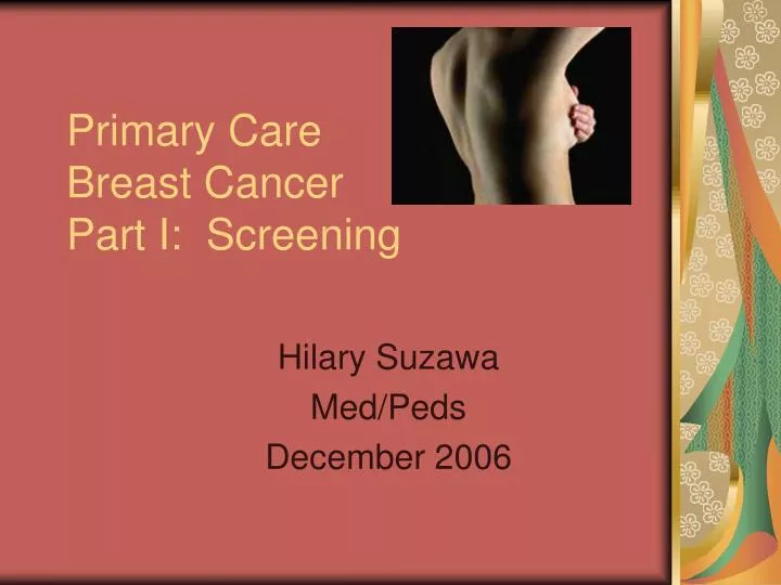
Primary Care Breast Cancer Part I: Screening. Hilary Suzawa Med/Peds December 2006. Breast Cancer Incidence. One in Eight (12%) : Lifetime risk of developing invasive breast CA Breast CA is the second most commonly dx CA among women (1 st —skin CA) ~180,000 new cases annually

E N D
Primary CareBreast CancerPart I: Screening Hilary Suzawa Med/Peds December 2006
Breast Cancer Incidence • One in Eight (12%): Lifetime risk of developing invasive breast CA • Breast CA is the second most commonly dx CA among women (1st—skin CA) • ~180,000 new cases annually • Estimated 212,920 dx in 2006 per Up To Date • Invasive breast CA accounts for 32% of new CA cases in American women • Pre-invasive breast CA (DCIS) now accounts for ~25-30% of all newly dx breast CA detected by mammogram
Breast Cancer Mortality • Second leading cause of cancer death in women (1st—lung CA) • ~48,000 deaths per year • Estimated 40,970 deaths in 2006 per Up To Date • Invasive breast CA accounts for 15% of CA deaths in American women • Main cause of death in women age 45-55 years • Annual mortality rates from breast CA have decreased over the last decade
Risk Factors • Most women with breast CA have no identifiable risk factors • Female • 1% of breast CA occur in men • Incidence of breast CA increases with age
Risk Factors • Race • Overall White > Black • Women <50 years: Black > White • Black women more likely to die of breast CA than white women • High social economic status
Risk Factors • Personal history of breast CA • Increases the risk of developing a new breast CA by 0.5-1% per year • Early menarche and late menopause—increased exposure to hormones • Give birth to their first child after age 30 or who never become pregnant
Risk Factors • OCP (?) • OCP <10 years has same risk as women who have never been on OCP • FMH: First degree relative, esp if dx pre-menopause • 3-4 x increase risk • Exposure to ionizing radiation • Eg. Survivors of Hodgkin’s Disease
????? • Chemical exposure • Alcohol consumption • Weight gain • High-fat diet • Induced abortion • Physical activity
Risk Reduction • Low fat, high fiber diet • Reduced alcohol consumption • Tamoxifen, Raloxifene
Genetics • ~8% of all cases of breast CA are hereditary • ~50% of these CA are related to BRCA-1 and BRCA-2 • Pre-menopausal women • Bilateral breast cancer
Genetic Screening • Insurance • Job discrimination • Prophylactic mastectomy and/or oophorectomy • False-negative tests
Clinical Presentations • Asymptomatic (screening only) • Breast Mass • Most common complaint • ~90% of all breast masses are benign • Fibroadenoma, Cyst • Breast Pain • Mastalgia is rarely assoc with breast CA • More common with fibrocystic change, HRT
Clinical Presentations • Skin changes • Erythema, edema, retraction of the skin or nipple • Nipple discharge • Discharge associated with Breast CA • Discharge is spontaneous • Assoc with a mass • Localized to a single duct in one breast
Clinical Breast ExamRecommendations • Part of Well Woman Exam • ACS recommendations • Pt age 20-39 years should have one every 3 years • Pt age 40 years and older annually
Clinical Breast Exam Key Points • Sitting and Supine positions • Differences in size (asymmetry) • Different arm positions • Retraction of skin or nipple • Prominent venous patterns • Signs of inflammation or skin changes (peau d’orange) • Nipple discharge • Axillary and supraclavicular LAD • Teach self-breast exam
Suspicious Findings • Mass • Solitary • Discrete • Hard • Fixed to skin or muscle (non-mobile) • Unilateral • Non-tender • Area of skin thickening • Breast CA is rarely bilateral when first dx/detected
Self Breast ExamsRecommendations • ACS (American Cancer Society) recommends start at age 20 years • Teach adolescents • Monthly • Same time each month, eg. Week after menses
MammogramRecommendations • American Cancer Society (ACS) and National Cancer Institute (NCI) • For asymptomatic women • Start age age 40 years • Annually • Screening women 50-75 years significantly decreases the death rate from breast CA • Screening women >75 years controversial but at any age screening detects breast CA at an earlier stage (risk-benefit analysis)
Mammogram Bottom Line: Uncomfortable but Necessary and Important
Mammogram False + • Women age 40-69 years have a 30% chance of false-positive screening mammogram OR breast exam over a 10-year period • False positive screening tests are higher for younger women because fewer of their breast masses are malignant (prevalence)
Mammogram False - • 10-15% of all breast CA are NOT detected by mammogram • A PALPABLE breast mass that is NOT seen on a mammogram should have a diagnostic work-up • Breast ultrasound • Needle biopsy • Close follow-up
Screening Recommendations Review • American Cancer Society • Age 20-39 years • Clinical breast exam every 3 years • Monthly self breast exam • Age 40 years and older • Annual mammogram • Annual clinical breast exam • Monthly self breast exam
Screening Recommendations • U.S. Preventive Services Task Force (USPSTF) • Routine screening in women for breast CA every 1-2 years • Mammography alone OR mammography and annual clinical breast exam for women age 50-69 years
Early Screening • Women with FMH of BRCA mutation should begin annual mammography between age 30-35 years • H/o chest radiation (XRT) • Mammograms may start as early as when patient age 20’s. • eg. h/o Hodgkin’s Disease: Children’s Oncology Group (COG) recommends start mammogram 8-10 years after chest XRT or at age 25 years (whichever later)
Other Imaging • Ultrasound • To differentiate b/t solid and cystic breast mass • Helpful in younger pt with dense breast tissue • Digital Mammogram • Images may be enhanced by modifying brightness or contrast • Initial studies show that digital mammograms are as accurate as standard radiographs • Not FDA approved
Evaluation of Common Problems • Cysts • Solid Masses • Nipple Discharge • Breast Pain • Pregnancy
Cysts • Ultrasound • Simple cyst • Round or oval • Sharp margins • Lacks internal echoes • Posterior acoustic enhancement • Simple vs. Complex Cyst • Aspiration of simple cyst • Evaluate any masses that remain after cyst aspirated
Solid Masses • Clinically suspicious mass should be followed even if normal mammogram • Ultrasound • FNA biopsy • Lumpectomy with 1-cm margin • Thickened area monitoring
Nipple Discharge • Suspicious for CA: spontaneous, assoc mass, single duct, bloody • Galactorrhea— • evaluate for prolactinoma • Cytology of discharge rarely helpful • Check mammogram • Ductogram
Breast Pain • Most commonly with fibrocystic change and uncommon with breast CA • Breast Exam +/- mammogram • Tx for fibrocystic breast dz • Pain meds • Firm support bra • Eliminate chocolate, caffeine • Vitamin E supplements
Pregnancy • Any mass in a pregnant or lactating woman should be thoroughly evaluated. • ~2% of breast CA are dx in pregnant women • Ultrasound • FNA biopsy and cytology
Susan G. Komen Foundation • Website at www.komen.org • Houston Affiliate • 713-783-9188 • Race for the Cure • Houston, TX • Planned for Saturday 10/6/2007 • Any Med/Peds runners?
Gifts that Matter • Consider purchasing holiday gifts that benefit Breast CA organizations • Susan G. Komen • Ford “Warriors in Pink” • Pink Ribbon Store at www.TheBreastCancerSite.com • Beauty Suppliers: Sephora, Origins
Bibliography • Apantaku L, “Breast Cancer Diagnosis and Screening.” American Family Physician 2000; 62 (3). • Hurria A, Joyce R, Come S, “Follow-up for breast cancer survivors: Patterns of relapse and long-term complications of therapy,” 3/10/06, Up To Date • Hurria A, Joyce R, Come S, “Follow-up for breast cancer survivors: Recommendations for surveillance after therapy,” 5/11/06, Up To Date • Esserman L and Stomper P,” Diagnostic evaluation and initial staging work-up of women with suspected breast cancer,” 3/29/06, Up To Date • Children’s Oncology Group Long-Term Follow-Up Guidelines at www.survivorshipguidelines.org
Primary CareBreast CancerPart II: Diagnosis, Treatment, Survivor Follow-up Hilary Suzawa Med/Peds February 2007
Overview • Palpable Mass • Imaging • Mammogram • Ultrasound • MRI • Breast Biopsy • Prognosis • Treatment Complications • Breast CA Survivor Follow-up • Recurrence vs. Second Primary
Breast Mass • If the lesion is palpable and the estimated likelihood of malignancy is >50%, then effort should be made to have surgeon evaluate prior to any biopsy procedure • Biopsy may lead to hematoma and inflammation (confounding)
Mammogram • Breast Exam alone NOT sufficient for breast CA diagnosis • Breast Cancer Detection Demonstration Project (BCDDP) • <10% of breast CA were detected solely by physical exam • >90% were identified by mammogram
Mammogram Bottom Line: Uncomfortable but Necessary and Important
MammogramScreening vs. Diagnostic • For women with sx or signs of breast CA, diagnostic mammogram is associated with higher sensitivity but lower specificity than screening mammogram • Note: • Sensitivity (Rule Out—“Snout”) • Specificity (Rule In–“Spin”)
MammogramViews and ACR Bi-RADS • Mammogram views • Spot compression • Magnification views • Varied angled views • ACR BI-RADS scale: American College of Radiology Breast Imaging Reporting and Data System
MammogramAbnormal Findings • 2 general categories of mammogram findings suggestive of breast CA • Soft tissue masses • Clustered micro-calcifications • Most specific mammographic feature of malignancy is spiculated soft tissue mass • Nearly 90% of these lesions represent invasive CA
MammogramMicro-calcifications • Micro-calcifications are seen in ~60% of CA detected by mammogram • Micro-calcifications are thought to represent intra-ductal calcification in areas of necrotic tumor • Mammogram appearance alone can NOT differentiate between purely intra-ductal and invasive ductal breast CA • ie, there is NO mammogram feature of basement membrane invasion
MammogramStaging • Multi-focal—several areas within one breast quadrant • Signifies disease along an entire duct • Multi-centric—multiple areas within different breast quadrants • Signifies involvement of multiple ducts • Intra-mammary LN mets • Worse prognosis
Breast Ultrasound • Adjunct to mammogram • To differentiate between solid and cystic masses • Negative predictive value in a patient with palpable breast mass and a non-suspicious mammogram is high (>99%) • Simple cysts need no further intervention because risk of CA is very low
Breast MRI • Nearly all breast invasive CA enhance on gadolinium contrast-enhanced MRI • Possible uses • Clinical staging • Screening of contra-lateral breast • Evolving role
Breast Biopsy • If pt has suspicious mammogram OR palpable mass, then biopsy • Percutaneous FNA • Percutaneous core needle biopsy • Vacuum-assisted biopsy • Wire localization and excision
Breast Biopsy • Fine-needle aspiration (FNA) • 20-gauge needle for sample from solid mass for cytology • Ultrasound or stereo-tactic guidance to assist in collecting FNA from a non-palpable mass • Core Biopsy • 14-gauge needle to remove cores of tissue from a mass • Ultrasound or stereo-tactic guidance • Small skin incision and local anesthesia
Breast Biopsy • Excisional Biopsy • May be the initial procedure of choice if high probability of malignancy • Wire localization of the mass if not palpable • Local anesthesia • May be done as outpatient