Download

1 / 64
841 likes | 1.49k Views
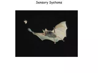
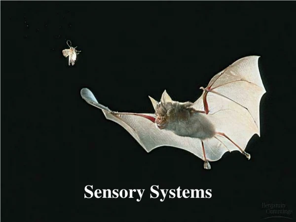
Sensory Systems. Mechanoreceptors. Transform mechanical stimuli into electrical signals All organisms (and most cells) sense and respond to mechanical stimuli Two main types of mechanoreceptor proteins: ENaC Epithelial sodium channels TRP channels Transient receptor potential channels

E N D
Mechanoreceptors • Transform mechanical stimuli into electrical signals • All organisms (and most cells) sense and respond to mechanical stimuli • Two main types of mechanoreceptor proteins: • ENaC • Epithelial sodium channels • TRP channels • Transient receptor potential channels • Channels are linked to extracellular matrix • Mechanical stimuli alter channel permeability
Touch and Pressure • Three classes of receptors • Baroreceptors • Interoceptors detect pressure changes • Tactile receptors • Exteroceptors detect touch, pressure, and vibration • Proprioceptors • Monitor the position of the body
Vertebrate Tactile Receptors • Widely dispersed in skin • Receptor structure • Free nerves endings • Nerve endings enclosed in accessory structures • (e.g., Pacinian corpuscle)
Vertebrate Proprioceptors • Monitor the position of the body • Three major groups • Muscle spindles • Located in skeletal muscles • Monitor muscle length • Golgi tendon organs • Located in tendons • Monitor tendon tension • Joint capsule receptors • Located in capsules that enclose joints • Monitor pressure, tension, and movement
Insect Tactile Receptors • Two common types of sensilla • Trichoid • Hairlike projection of cuticle • Bipolar sensory neuron • TRP channel • Campaniform • Dome-shaped bulge of cuticle • Bipolar sensory neuron
Insect Proprioceptors • Scolopidia • Bipolar neuron and complex accessory cell (scolopale) • Can be isolated or grouped into chordotonal organs • Most function in proprioception • Can be modified into tympanal organs for sound detection
Equilibrium and Hearing • Utilize mechanoreceptors • Equilibrium (“balance”) • Detect position of the body relative to gravity • Hearing • Detect and interpret sound waves • Vertebrates • Ear is responsible for equilibrium and hearing • Invertebrates • Organs for equilibrium are different from organs of hearing
Statocysts • Organ of equilibrium in invertebrates • Hollow, fluid filled cavities lined with mechanosensory neurons • Statocysts contain statoliths • Dense particles of calcium carbonate • Movement of statoliths stimulate mechanoreceptors
Insect Hearing • Strong vibrations sensed by trichoid sensilla • Weak vibrations and sounds are detected by chordotonal organs • Clusters of scolopidia • Located on leg • Mechanosensitive ion channels • Tympanal organs • Thin layer of cuticle (tympanum) overlays chordotonal organ
Vertebrate Hair cells • Mechanoreceptor for hearing and balance • Modified epithelial cells (not neurons) • Cilia on apical surface • Kinocilium (a true cilium) • Stereocilia (microvilli) • Tips of stereocilia are connected by proteins (tip links) • Mechanosensitive ion channels in stereocilia • Movement of stereocilia change in permeability • Change in membrane potential • Change in release of neurotransmitter from hair cell
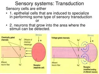
Signal Transduction in Hair Cells Figure 6.18
Fish and Amphibian Hair Cells • Hair cells detect body position and movement • Neuromast • Hair cells and cupula • Stereocilia embedded in gelatinous cap • Detect movement of water • Lateral line system • Array of neuromasts within pits or tubes running along the side of the body Fish Neuromast
Vertebrate Ears • Function in both equilibrium and hearing • Outer ear • Not in all vertebrates • Pinna • Auditory canal • Middle ear • Not in all vertebrates • Interconnected bones in air-filled cavity • Inner ear • Present in all vertebrates • Series of fluid-filled membranous sacs and canals • Contains mechanoreceptors (hair cells) Mammalian Ear
Inner Ear: Vestibular Apparatus and Cochlea • Vestibular apparatus detects movements • Three semi-circular canals with enlarged region at one end (ampulla) • Two sacklike swellings (utricle and saccule) • Lagena • Extension of saccule • Extended in birds and mammals into a cochlear duct or cochlea for hearing • Hair cells present in vestibular apparatus and lagena (cochlea)
Vestibular Apparatus (1) • Mechanoreceptors of the inner ear • Macula • Present in utricle and saccule • Mineralized otoliths suspended in a gelatinous matrix • Stereocilia of hair cells embedded in matrix • >100,000 hair cells • Detect linear acceleration and tilting of head
Vestibular Apparatus (2) • Cristae • Present in ampullae of semicircular canals • Gelatinous matrix (cupula) lacks otoliths • Stereocilia of hair cells embedded in matrix • Detect angular acceleration (turning) of head
Maculae Detect Linear Acceleration and Tilting Figure 6.23
Cristae Detect Angular Acceleration Figure 6.24
Sound Detection by Inner Ear • Fish • Sound waves cause otoliths to move • Displacement of cilia on hair cells • Some fish use the swim bladder to amplify sounds Figure 6.25
Sound Detection by Inner Ear • Terrestrial Vertebrates • Hearing involves the inner, middle, and outer ear • Sound transfers poorly between air and the fluid-filled inner ear • Amplification of sound waves • Pinna acts as a funnel to collect more sound • Middle ear bones increase the amplitude of vibrations from the tympanic membrane to the oval window
Mammalian Middle and Inner Ear Figure 6.26a
Mammalian Inner Ear • Specialized for sound detection • Perilymph • Fills vestibular and tympanic ducts • Similar to extracellular fluids (high Na+ and low K+) • Endolymph • Fills cochlear duct • Different from extracellular fluid (high K+ and low Na+) • Organ of Corti • Hair cells on basilar membrane • Inner and outer rows of hair cells • Stereocilia embedded in tectorial membrane in cochlear duct (filled with endolymph)
Mammalian Inner Ear Figure 6.26a,b
Sound Transduction • Sound waves vibrate tympanic membrane • Middle ear bones transmit vibration to oval window • Oval window vibrates • Pressure waves in perilymph of vestibular duct • Basilar membrane vibrates • Stereocilia on the inner hair cells bend • Hair cells depolarize • Hair cells release neurotransmitter (glutamate) • Glutamate excites sensory neuron • Round window serves as a pressure valve
Encoding Sound Frequency • Frequency Detection • Basilar membrane is stiff and narrow at the proximal end and flexible and wide at distal end • High frequency sound vibrates stiff end • Low frequency sound vibrates flexible end • Specific regions of brain respond to specific frequencies • Place coding
Encoding Sound Amplitude and Amplification • Amplitude Detection • Loud sounds cause larger movement of basilar membrane than quiet sounds • depolarization of inner hair cells • AP frequency • Outer hair cells amplify quiet sounds • Change shape in response to sound • Do not release neurotransmitter • Change in shape increases movement of basilar membrane • Increased stimulus to inner hair cells
Detecting Sound Location • Brain uses time lags and differences in sound intensity to detect location of sound • Sound in right ear first • Sound located to the right • Sound louder in right ear • Sound located to the right • Rotation of head helps localize sound
Photoreception • Ability to detect visible light • A small proportion of the electromagnetic spectrum from ultraviolet to near infrared • Ability to detect this range of wavelengths supports idea that animals evolved in water • Visible light travels well in water; other wavelengths do not
Electromagnetic Spectrum Figure 6.27a,b
Photoreceptors • Range from single light-sensitive cells to complex, image-forming eyes • Two major types of photoreceptor cells: • Ciliary photoreceptors • Have a single, highly folded cilium • Folds form disks that contain photopigments • Rhabdomeric photoreceptors • Apical surface covered with multiple outfoldings called microvillar projections • Microvillar projections contain photopigments • Photopigments • Molecules that absorb energy from photons
Phylogeny of Photoreceptors Figure 6.28
Vertebrate Photoreceptors • Vertebrates have ciliary photoreceptors • Rods • Cones • Both have inner and outer segments • Inner and outer segments connected by a cilium • Outer segment contains photopigments • Inner segment forms synapses with other cells
Characteristics of Rods and Cones Table 6.1
Diversity in Rod and Cone Shape • Diverse shapes of rods and cones among vertebrates • Shape does not determine properties of photoreceptor • Properties of photoreceptor depend on its photopigment Figure 6.30
Photopigments • Photopigments have two covalently bonded parts • Chromophore • Derivative of vitamin A • For example, retinal • Contains carbon-carbon double bonds • Absorption of light converts bond from cis to trans • Opsin • G-protein-coupled receptor protein • Opsin structure determines photopigment characteristics • For example, wavelength of light absorbed
Phototransduction • Steps in photoreception • Chromophore absorbs energy from photon • Chromophore changes shape • Double bond isomerizes from cis to trans • Activated chromophore dissociates from opsin • “Bleaching” • Opsin activates G-protein • Formation of second messenger • Ion channels open or close • Change in membrane potential
Phototransduction Figure 6.32
The Eye • Eyespots • Cells or regions of a cell that contain photosensitive pigment • For example, protist Euglena • Eyes are complex organs • Detect direction of light • Light-dark contrast • Some can form an image
Types of Eyes • Flat sheet eyes • Some sense of light direction and intensity • Often in larval forms or as accessory eyes in adults Figure 6.33a
Types of Eyes • Cup-shaped eyes (e.g., Nautilus) • Retinal sheet is folded to form a narrow aperture • Discrimination of light direction and intensity • Light-dark contrast • Image formation • Poor resolution Figure 6.33b
Types of Eyes • Vesicular Eyes (present in most vertebrates) • Lens in the aperture improves clarity and intensity • Lens refracts light and focuses it onto a single point on the retina • Image formation • Good resolution Figure 6.33c
Types of Eyes • Convex Eye (annelids, molluscs, arthropods) • Photoreceptors radiate outward • Convex retina Figure 6.33d
Compound Eyes of Arthropods • Composed of ommatidia (photoreceptor) • Each ommatidium has its own lens • Images formed in two ways • Apposition compound eyes • Ommatidia operate independently • Each one detects only part of the image • Afferent neurons interconnect to form an image • Superposition compound eyes • Ommatidia work together to form image • Resolving power is increased by reducing size and increasing the number of ommatidia
Structure of The Vertebrate Eye • Sclera • “White” of the eye • Cornea • Transparent layer on anterior • Retina • Layer of photoreceptor cells • Choroid • Pigmented layer behind retina • Tapetum • Layer in the choroid of nocturnal animals that reflects light
Structure of the Vertebrate Eye • Iris • Two layers of pigmented smooth muscle • Pupil • Opening in iris allows light into eye • Lens • Focuses image on retina • Ciliary body • Muscles that change lens shape • Aqueous humor • Fluid in the anterior chamber • Vitreous humor • Gelatinous mass in the posterior chamber
Image Formation • Refraction – bending of light rays • Cornea and lens focus light on the retina • In terrestrial vertebrates, most of the refraction occurs between air and cornea • Lens does fine focusing • Lens changes shape to focus on near or far objects • Accommodation
Image Accommodation • Accommodation • Light rays must converge on the retina to produce a clear image • Focal point • Point at which light waves converge • Focal distance • Distance from a lens to its focal point
Image Accommodation • Distant objects • Light rays are parallel when entering the lens • Ciliary muscles contract • Lens is pulled and becomes thinner • Little refraction of light by lens • Close objects • Light rays are not parallel when entering the lens • Ciliary muscles relax • Lens becomes thicker • More refraction of light by lens
Image Accommodation Figure 6.36