Download
1 / 58
580 likes | 599 Views
Understand DNA's double helix structure, replication process, contributions to science by key figures, and the molecular basis of inheritance, including transformation and DNA as genetic material. Learn about nucleotide composition, Chargaff's Rules, function of enzymes involved in DNA replication, and the significance of telomeres. Compare prokaryotic and eukaryotic DNA, replication models, and the Meselson-Stahl experiment proving DNA replication is semi-conservative. Enhance your knowledge for a comprehensive understanding of DNA.

E N D
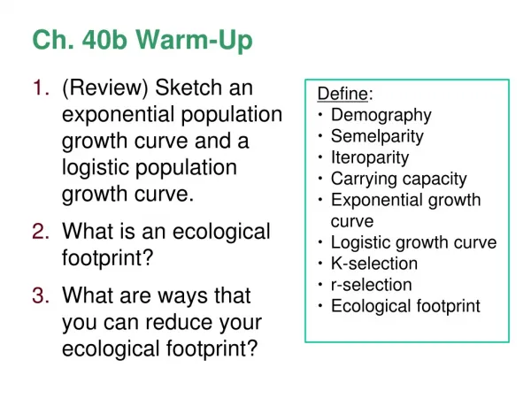
Ch. 16 Warm-Up • Draw and label a nucleotide. • Why is DNA a double helix? • What is the complementary DNA strand to: DNA: A T C C G T A T G A A C
Ch. 16 Warm-Up • What was the contribution made to science by these people: • Hershey and Chase • Franklin • Watson and Crick • Chargaff’s Rules: If cytosine makes up 22% of the nucleotides, then adenine would make up ___ % ? • Explain the semiconservative model of DNA replication.
Ch. 16 Warm-Up • What is the function of the following: • Helicase • DNA Ligase • DNA Polymerase (I and III) • Primase • Nuclease • How does DNA solve the problem of slow replication on the lagging strand? • Code the complementary DNA strand: 3’ T A G C T A A G C T A C 5’ • What is the function of telomeres?
THE MOLECULAR BASIS OF INHERITANCE Chapter 16
What you must know • The structure of DNA. • The major steps to replication. • The difference between replication, transcription, and translation. • The general differences between the bacterial chromosome and eukaryotic chromosomes. • How DNA is packaged into a chromosome.
Problem: Is the genetic material of organisms made of DNA or proteins?
Frederick Griffith (1928) S strain of bacteria = pathogenic (causes pneumonia in mice) R strain of bacteria = non-pathogenic HOW?
The “Transforming Principle” mix heat-killed pathogenic & non-pathogenic bacteria live pathogenic strain of bacteria live non-pathogenic strain of bacteria heat-killed pathogenicbacteria A. B. D. C. mice die mice live mice live mice die Transformation=change in phenotype something in heat-killed bacteria could still transmit disease-causing properties
Frederick Griffith (1928) Conclusion: living R bacteria transformed into deadly S bacteria by an unknown, heritable substance (R bacteria must have acquired something from the S bacteria) - Still unclear WHAT the transforming, heritable substance was…until… Oswald Avery, et al. (1944) • Discovered that the transforming agent was DNA… But still skepticism from scientific community
Hershey and Chase (1952) • Bacteriophages: virus that infects bacteria; composed of DNA and protein To answer the question: Is DNA or PROTEIN the heritable substance that determines traits? Protein = radiolabel S (radioactive sulfur label) DNA = radiolabel P (radioactive phosphorus label)
Hershey & Chase Protein coat labeled with 35S DNA labeled with 32P T2 bacteriophages are labeled with radioactive isotopes S vs. P bacteriophages infect bacterial cells bacterial cells are agitated to remove viral protein coats Which radioactive marker is found inside the cell? Which molecule carries viral genetic info? 32P radioactivity foundin the bacterial cells 35S radioactivity found in the medium
Hershey and Chase (1952) Conclusion: DNA entered infected bacteria DNA must be the genetic material!
Hershey & Chase 1952 | 1969 Hershey Martha Chase Alfred Hershey
Edwin Chargaff (1947) Chargaff’s Rules: • DNA composition varies between species • Ratios: • %A = %T and • %G = %C
Rosalind Franklin (1950’s) • Worked with Maurice Wilkins • X-ray crystallography = images of DNA • Provided measurements on chemistry of DNA
James Watson & Francis Crick (1953) • Discovered the double helix by building models to conform to Franklin’s X-ray data and Chargaff’s Rules.
1953 article in Nature Watson and Crick Watson Crick
Structure of DNA DNA = double helix • “Backbone” = sugar + phosphate • “Rungs” = nitrogenous bases
Structure of DNA Nitrogenous Bases • Adenine (A) • Guanine (G) • Thymine (T) • Cytosine (C) • Pairing: • purine + pyrimidine • A = T • G Ξ C purine pyrimidine
Structure of DNA Hydrogen bondsbetween base pairs of the two strands hold the molecule together like a zipper.
Structure of DNA Antiparallel: one strand (5’ 3’), other strand runs in opposite, upside-down direction (3’ 5’)
DNA Comparison Prokaryotic DNA Eukaryotic DNA • Double-stranded • Circular • One chromosome • In cytoplasm • No histones • Supercoiled DNA • Double-stranded • Linear • Usually 1+ chromosomes • In nucleus • DNA wrapped around histones (proteins) • Forms chromatin
Problem: How does DNA replicate?
Replication: Making DNA from existing DNA 3 alternative models of DNA replication Newly made DNA is light blue
Pulse-Chase Primer: The Meselson-Stahl Experiment • Read the Introduction
DNA Replication Video Amoeba Sisters – with handout
Major Steps of Replication: • Helicase:unwinds DNA at origins of replication • Initiation proteins separate 2 strands forms replication bubble • Primase: puts down RNA primer to start replication • DNA polymerase III: adds complimentary bases to leading strand (new DNA is made 5’ 3’) • Lagging strand grows in 3’5’ direction by the addition of Okazaki fragments • DNA polymerase I: replaces RNA primers with DNA • DNA ligase: seals fragments together
1. Helicase unwinds DNA at origins of replication and creates replication forks
4. DNA polymerase III adds nucleotides in 5’3’ direction on leading strand
Okazaki Fragments: Short segments of DNA that grow 5’3’ that are added onto the Lagging Strand DNA Ligase: seals together fragments