Download
1 / 33
360 likes | 650 Views
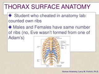
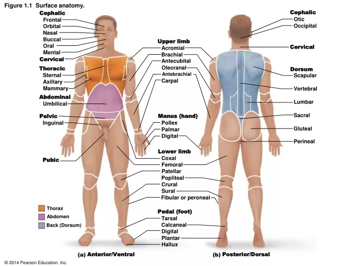
Cephalic. Cephalic. Otic. Frontal. Occipital. Orbital. Nasal. Buccal. Upper limb. Oral. Cervical. Acromial. Mental. Brachial. Cervical. Antecubital. Olecranal. Thoracic. Dorsum. Antebrachial. Sternal. Scapular. Carpal. Axillary. Figure 1.1 Surface anatomy. Mammary.

E N D
Cephalic Cephalic Otic Frontal Occipital Orbital Nasal Buccal Upper limb Oral Cervical Acromial Mental Brachial Cervical Antecubital Olecranal Thoracic Dorsum Antebrachial Sternal Scapular Carpal Axillary Figure 1.1 Surface anatomy. Mammary Vertebral Abdominal Lumbar Umbilical Sacral Manus (hand) Pelvic Pollex Inguinal Gluteal Palmar Digital Perineal Lower limb Coxal Pubic Femoral Patellar Popliteal Crural Sural Fibular or peroneal Thorax Pedal (foot) Abdomen Tarsal Calcaneal Back (Dorsum) Digital Plantar Hallux Anterior/Ventral Posterior/Dorsal
Figure 1.1a Surface anatomy. Cephalic Frontal Orbital Nasal Buccal Upper limb Oral Acromial Mental Brachial Cervical Antecubital Olecranal Thoracic Antebrachial Sternal Carpal Axillary Mammary Abdominal Umbilical Manus (hand) Pelvic Pollex Inguinal Palmar Digital Lower limb Coxal Pubic Femoral Patellar Crural Fibular or peroneal Thorax Pedal (foot) Abdomen Tarsal Back (Dorsum) Digital Hallux Anterior/Ventral
Figure 1.1b Surface anatomy. Cephalic Otic Occipital Upper limb Cervical Acromial Brachial Olecranal Dorsum Antebrachial Scapular Vertebral Lumbar Sacral Manus (hand) Gluteal Digital Perineal Lower limb Femoral Popliteal Sural Fibular or peroneal Thorax Abdomen Pedal (foot) Back (Dorsum) Calcaneal Plantar Posterior/Dorsal
Superior (cephalad) Figure 1.2 Anatomical terminology describing body orientation and direction. Posterior (dorsal) Anterior (ventral) Superior (dorsal) Proximal Posterior (caudal) Anterior (cephalad) Distal Inferior (ventral) Inferior (caudal)
Figure 1.2a Anatomical terminology describing body orientation and direction. Superior (cephalad) Posterior (dorsal) Anterior (ventral) Proximal Distal Inferior (caudal)
Figure 1.2b Anatomical terminology describing body orientation and direction. Superior (dorsal) Posterior (caudal) Anterior (cephalad) Inferior (ventral)
Figure 1.3 Planes of the body with corresponding magnetic resonance imaging (MRI) scans. Frontal plane Median (midsagittal) plane Transverse plane Transverse section (through torso, inferior view) Median (midsagittal) section Frontal section (through torso) Spinal cord Pancreas Left and right lungs Liver Heart Liver Intestines Vertebral column Aorta Rectum Spleen Arm Subcutaneous fat layer Body wall Stomach
Figure 1.3a Planes of the body with corresponding magnetic resonance imaging (MRI) scans. Frontal plane Median (midsagittal) plane Transverse plane Frontal section (through torso) Left and right lungs Liver Heart Arm Stomach
Figure 1.3b Planes of the body with corresponding magnetic resonance imaging (MRI) scans. Frontal plane Median (midsagittal) plane Transverse plane Transverse section (through torso, inferior view) Spinal cord Pancreas Liver Aorta Spleen Subcutaneous fat layer Body wall
Figure 1.3c Planes of the body with corresponding magnetic resonance imaging (MRI) scans. Frontal plane Median (midsagittal) plane Transverse plane Median (midsagittal) section Intestines Vertebral column Rectum
Figure 1.4 Objects can look odd when viewed in section. Cross section Midsagittal section Frontal sections
Figure 1.4a Objects can look odd when viewed in section. Cross section
Figure 1.4b Objects can look odd when viewed in section. Midsagittal section
Figure 1.4c Objects can look odd when viewed in section. Frontal sections
Figure 1.5 Dorsal and ventral body cavities and their subdivisions. Cranial cavity Cranial cavity (contains brain) Vertebral cavity Superior mediastinum Dorsal body cavity Thoracic cavity (contains heart and lungs) Pleural cavity Pericardial cavity within the mediastinum Vertebral cavity (contains spinal cord) Ventral body cavity (thoracic and abdominopelvic cavities) Diaphragm Abdominal cavity (contains digestive viscera) Abdomino- pelvic cavity Pelvic cavity (contains urinary bladder, reproductive organs, and rectum) Dorsal body cavity Ventral body cavity Anterior view Lateral view
Figure 1.5a Dorsal and ventral body cavities and their subdivisions. Cranial cavity (contains brain) Superior mediastinum Dorsal body cavity Thoracic cavity (contains heart and lungs) Pleural cavity Pericardial cavity within the mediastinum Vertebral cavity (contains spinal cord) Diaphragm Abdominal cavity (contains digestive viscera) Pelvic cavity (contains urinary bladder, reproductive organs, and rectum) Dorsal body cavity Ventral body cavity Lateral view
Figure 1.5b Dorsal and ventral body cavities and their subdivisions. Cranial cavity Vertebral cavity Superior mediastinum Thoracic cavity (contains heart and lungs) Pleural cavity Pericardial cavity within the mediastinum Ventral body cavity (thoracic and abdominopelvic cavities) Diaphragm Abdominal cavity (contains digestive viscera) Abdomino- pelvic cavity Pelvic cavity (contains urinary bladder, reproductive organs, and rectum) Dorsal body cavity Ventral body cavity Anterior view
Figure 1.6 Serous membranes of the ventral body cavities. Parietal peritoneum Parietal pleura Visceral peritoneum Visceral pleura Parietal pericardium Visceral pericardium
Figure 1.6 Serous membranes of the ventral body cavities. (1 of 2) Parietal peritoneum Visceral peritoneum
Figure 1.6 Serous membranes of the ventral body cavities. (2 of 2) Parietal pleura Visceral pleura Parietal pericardium Visceral pericardium
Figure 1.7 Abdominopelvic quadrants. Right upper quadrant (RUQ) Left upper quadrant (LUQ) Right lower quadrant (RLQ) Left lower quadrant (LLQ)
Figure 1.8 Abdominopelvic regions. Diaphragm Liver Right hypochondriac region Left hypochondriac region Epigastric region Spleen Gallbladder Stomach Transverse colon of large intestine Ascending colon of large intestine Right lumbar region Left lumbar region Umbilical region Descending colon of large intestine Small intestine Cecum Initial part of sigmoid colon Left iliac (inguinal) region Right iliac (inguinal) region Hypogastric (pubic) region Appendix Urinary bladder
Figure 1.8a Abdominopelvic regions. Right hypochondriac region Left hypochondriac region Epigastric region Right lumbar region Left lumbar region Umbilical region Hypogastric (pubic) region Left iliac (inguinal) region Right iliac (inguinal) region
Figure 1.8b Abdominopelvic regions. Diaphragm Liver Spleen Stomach Gallbladder Transverse colon of large intestine Ascending colon of large intestine Descending colon of large intestine Small intestine Cecum Initial part of sigmoid colon Appendix Urinary bladder
Figure 1.9 Other body cavities. Middle ear cavity Orbital cavity (orbit) Synovial cavity in a joint between neck vertebrae Nasal cavity Fibrous layer around joint Oral cavity (mouth) Tongue
Review Figure 1.5 Body cavities 3 cavity 1 Dorsal body cavity (superior) 4 cavity (inferior) 5 cavity 2 Ventral body cavity (superior) 7 cavity cavity 6 (inferior) (superior) cavity 8 (inferior)