Download
1 / 26
260 likes | 278 Views
Learn about SVC syndrome, its causes, symptoms, and radiographic studies. Discover treatment options including radiotherapy, medical management, and surgical interventions.

E N D
Radiotherapy for SVC syndrome SomvilaiChakrabandhu, MD. Division of Therapeutic Radiology and Oncology, Faculty of Medicine, Chiang Mai University
SVC syndrome Definition • The clinical manifestation of obstruction of the superior vena cava, with severe reduction in venous return from the head, neck, and upper extremities
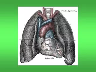
SVC syndrome Superior vena cava • carries venous blood from the head, arms, and upper trunk to the heart • carries approximately one third of the venous return to the heart.
SVC syndrome Obstruction of Superior vena cava • Reduction in venous return of face, neck, upper extremities • Collateral development of venous system - azygos, internal thoracic, paraspinous, esophageal
SVC syndrome Types of SVC obstruction • Extrinsic compression • Mediastinal structure or lymph nodes • Intrinsic obstruction • Thrombosis • Neoplastic infiltration
SVC syndrome Causes of SVC obstruction • Malignant about 80% • Lung cancer • Lymphoma • Metastasis malignancy to mediastinal LNs • Non-malignancy • Infection (stphilis, TB) • Fibrosis • Thrombus (central venous catheter)
SVC syndrome • Dyspnea (most common) • Orthopnea • Facial / Neck swelling • Cough • hoarseness • Headache • Nasal congestion • Hemoptysis • Dysphagia • Dizziness • Syncope Symptoms
SVC syndrome • The severity of the symptoms depends on the degree of narrowing of the superior vena cava • Symptom onset depends on speed of SVC obstruction onset • Malignant disease can arise in weeks to months • Not enough time to develop collaterals
SVC syndrome Physical finding • Edema of face, arms • Dilated neck veins • Increased collateral veins over anterior chest wall • Cyanosis • Severe cases include proptosis, glossal and laryngeal edema
Radiographic Studies Chest x-ray • Most common findings • Mediastinal widening • Pleural effusion
Radiographic Studies • CT Chest with contrast • Preferred choice • defines the level of obstruction • Maps out collateral pathways • Can differentiate between vena caval thrombosis and extrinsic compression
Radiographic Studies CT scan : Diagnosis level of obstruction
Radiographic Studies MRI • useful in patients with IV contrast allergies Positronemission tomography (PET) • sometime useful
SVC syndrome Management • Superior vena cava syndrome associated with malignant conditions involves both • treatment of the cancer and • relief of the symptoms of obstruction
SVC syndrome Emergency condition • Stridor from laryngeal edema and impending airway obstruction • Confusion related to associated cerebral edema Immediate action is needed • Attention to the ABCs assessment • Stabilize the airway
SVC syndrome Non- Emergency condition • Most patients are not in immediate danger at presentation • Sit upright : relief of the usual dyspnea • Oxygen support, if indicated • Consider steroids
SVC syndrome Treatments & interventions Medical management : • Corticosteroid and diuretic for laryngeal and cerebral edema (controversy) • Thrombolytic drug : thrombotic cause
SVC syndrome Treatments & interventions • Surgical treatment : bypass • Endovascular stent • increasingly used • immediate relief symptom • Refractory to RT/ chemotherapy
SVC syndrome Treatments & interventions • RT and chemotherapy • Relief symptom and • Treatment malignancy
Radiation Therapy • Excellent symptom relief: • dyspnea • edema of face and • distention of neck and thoracic vein • Symptomatic improvement usually takes 1-2 weeks afterradiotherapy
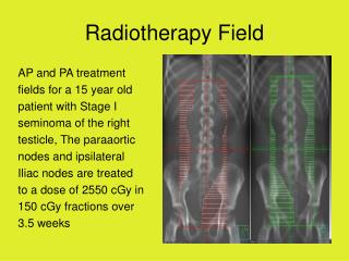
Radiation Therapy • Radiation dose for palliation • 20 - 30 Gy in 5 – 10 fractions • Depend on patient condition • Radiation field • Encompass mediastinal lymph nodes / hilar region
Radiation Therapy Supine position