Download
1 / 53
530 likes | 697 Views
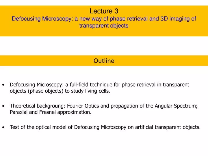
Lecture 3 Defocusing Microscopy: a new way of phase retrieval and 3D imaging of transparent objects. Outline. Defocusing Microscopy: a full-field technique for phase retrieval in transparent objects (phase objects) to study living cells.

E N D
Lecture 3 Defocusing Microscopy: a new way of phase retrieval and 3D imaging of transparent objects Outline • Defocusing Microscopy: a full-field technique for phase retrieval in transparent objects (phase objects) to study living cells. • Theoretical backgroung: Fourier Optics and propagation of the Angular Spectrum; Paraxial and Fresnel approximation. • Test of the optical model of Defocusing Microscopy on artificial transparent objects.
Motivation: Study of AdheredMacrophageMotility Film accelerated 16x
Phagocytosis of Leishmaniaamazonensis at 37oC Film accelerated 16x
Defocusingmicroscopy Adhered Macrophage Df = 0 Df > 0 Df < 0 Infinity corrected microscope Agero et al., PRE 67 (5), 051904 (2003) and Phys.Rev. Focus, May 21 (2003); Agero et al., Microsc. Res. Tech. 65, 159 (2004); Mesquita et al., APL (2006); Coelho-Neto et al., Biophysical J. (2006)
Light electric field for a defocused microscope Angular spectrum of the light electric field 2D Fourier transform Free propagation of the angular spectrum From Helmholtz equation propagating wave evanescent wave Considering a single polarization, propagation along z>0 and the paraxial approximation q<<k
Angular spectrum through a thin lens • From the object (z=0) to L1 (z=f1-∆f); • through L1 • from L1 to L2 (distance d) • through L2 • from L2 to the image plane I (distance f2 ) Electric field for the defocused microscope on the image plane
Diffraction by a sinusoidal phase grating with spacing L focal plane f light are Bessel functions of order m Electric field for the defocused sinusoidal phase grating
Contrast of a defocused phase grating considering only first order diffraction Defining contrast as with
SinusoidalPhaseGratingwithSpacing L Shifted Talbot Images
CurvedThickPhaseObject Objective Focal Position Glass-slide solution Light Z h(x,y) Df
PolystyreneSpherical Cap AFM image DM image
AFM and DM Profiles DM AFM R=5.12mm
RefractiveIndexDifferenceObtainedwith DM Dn=0.61±0.01
CCD Calibration Camera Dage-MTI – 8 bts Camera UniqVision – 12 bits Power meter intensity (mW)
Fluctuating transparent interfaces and contrast correlation function h Time average H with and for a stationary process such that Space-time correlation function of contrast fluctuations
Mean-square fluctuation of contrast and for the continuum case
Numerical example Mean square contrast fluctuation Spacial power spectrum of fluctuations
Diffraction by two transparent interfaces Average contrast Constrast correlation function
Numerical example Two symmetric interfaces Two asymmetric interfaces
Summary By using the propagation of the light angular spectrum we develop an optical model for a defocused bright-field microscope. Transparent objects can be visualized in a defocused microscope, since defocusing introduces a phase difference between the diffracted and transmitted light, which is translated into contrast after interference in the image plane. For small defocusing the average contrast of a surface is proportional to its curvature. We were able to obtain theoretical expressions for the correlation functions for one and two fluctuating interfaces. In the next lecture we will see, by using these expressions, how to obtain elastic information from the interfaces of living cells.
Lecture 4 Application of defocusing microscopy to study living cell motility Outline Application of the expressions obtained in Lecture 3 for testing motility models of living cells. Macrophages and phagocytocis: 3D imaging and study of fluctuations. Effects of nonequilibrium. Red Blood Cell: 3D imaging and study of coupling between the spectrin cytoskeleton and lipid bilayer via flickering. Effects of nonequilibrium.
ruffle SRMF Results – Curvature Fluctuations
Actin filaments just below the plasmatic membrane Cytoskeleton Polimerized protein filaments Alberts, et al Mol. Biol. Cell. 3rd Ed.Garland Pub. Inc. NY(1994) Svitkina, Verkhovsky, MacQuade & Borisy J. Cell Biol. 139 (2), 397 (1997)
Ruffles: curvature and thickness profiles Ruffle hyperbolic
Ruffles: curvature and thickness profiles Ruffle gaussian Measuring ruffle contrast as a function of defocusing we are able to obtain its refractive index. (Coelho Neto, Biophys. J. 91, 2006)
Time correlation function For bone marrow macrophages (extracted from healthy mice) this relaxation time is
Before and after addition of 100nM of Cytochalasin-D Ruffles are inhibited
Results: 24-37oC Coelho Neto et al., Exp. Cell Res. 303 (2), 207 (2005)
2d a f Actin filament b D x membrane actin-g Dynamics of actin polymerization (diffusion + polymerization) Discussion of models Model of cellular motility : Brownian Ratchet Peskin, Odell & Oster Biophys. J. 65, 316 (1993) Mogilner & Oster Biophys. J. 71, 3030 (1996) Mogilner & Oster Biophys. J. 84, 1591 (2003)
Phagocytosis of Leishmania amazonensis at 37oC Film accelerated 16x
Results: Phagocytosis at 37oC Behavior of <2> near the phagossome
Results: Phagocytosis from 24 to 37oC Coelho Neto et al., Exp. Cell Res. 303 (2), 207 (2005)
Protein-Membrane Coupling Model
Theoretical model of Experimental data of Coelho Neto et al. Exp. Cell Res. 303, 207 (2005)
RedBloodCell (RBC) Objective focal plane above the RBC middle plane Objective focal plane below the RBC middle plane Brochard – Lennon (1975), flickering due to thermal motion of surfaces
DefocusedImage of a RedBloodCell (RBC) Dn = 0.056 Mesquita, Agero, Mesquita, APL 88, 133901 (2006)
Limite assintótico para grandes desfocalizações Para Para hemácias
MembraneElasticityandFluctuations S. A. Safram, Statistical Thermodynamics of Surfaces, Interfaces, and Membranes, Addison-Wesley (1994). Membrane Free Energy Variation Monge representation u water bendingmodulus surfacetension Lipid bilayer Hydrophilic confinementpotential Fourier decomposition and energy equipartition Hydrophobic water Curvature energy for curved surfaces – Helfrich free-energy (Phys. Lett.1973) spontaneouscurvature maincurvatures Gaussiancurvature
d RBC ElasticModel of Auth, Safran, and Gov -Brochard F. and Lennon J.F., J. Physique , 36, 1035 (1975); -Zilker A., Engelhardt H., and Sackmann E., J. Physique 48, 2139 (1987); -Evans E., Methods Enzymol. 173, 3 (1989); -Tuvia S., Levin S. and Korenstein R, Proc Natl. Acad. Science, 94, 5045 (1997); -Tuvia S., Levin S. Bither A. and Korenstein R., J. Cell Biol. 141, 1551 (1998); -Gov N., Zilman A.G. and Safran S., Physical Review Letters 90 (22), 228101 (2003); -Gov N. and Safran S., Biophys. J. 88 (22), 1859 (2005); -Auth T., Safran S. and Gov N., Physical Review E 76 , 051910 (2007). Cytoskeleton is modeled as a hexagonal network of entropicsprings spectrin Spectrin filaments Actin nodes bilayer • B. Albertset al.,”Molecular • Biology of theCell”, (2002) ATP drivennon-thermaleffects Detached filaments
RBC ElasticModel of Auth, Safran, and Gov bilayer curvature modulus m cytoskeleton shear modulus effective temperature h cytoplasm viscosity
ReferenceSystem origen light Symmetry plane Glass-slide se sendo
Measurements of RBC Flickeringwith DM G. Glionna et al. APL (2009) Middle region of a RBC Defocusing microscopy is able to provide quantitative data about the fluctuations of each interface of a RBC separately. Contrast correlation between the same pixel after 33ms. The decay of large wavenumber fluctutations is evident in the figure.