Download
1 / 50
640 likes | 1.01k Views
Fibroids. Dr. Haresh U. Doshi M.D., Diploma (USG), FICOG, PGDMLS Professor & Chief of Unit B.J. Med. College , New Civil Hosp. Ahmedabad. Fibroid. Synonyms : Myoma, Leiomyoma, Fibromyoma Most common benign neoplasm in the female.

E N D
Dr. Haresh U. Doshi M.D., Diploma (USG), FICOG, PGDMLS Professor & Chief of Unit B.J. Med. College , New Civil Hosp. Ahmedabad
Fibroid • Synonyms : Myoma, Leiomyoma, Fibromyoma • Most common benign neoplasm in the female. • Incidence : 20 to 40% of reproductive age women.
Fibroid Etiology : It arises from smooth muscle cell of myometrium. • Exact etiology not known. • Monoclonal origin ( arising from single cell confirmed by G6PD studies. • Genetic basis definite. • Various growth factors like TGFβ , EGF, IGF-1, IGF-2, BFGF are recently implicated in the development of fibroids.
Fibroid - Etiology Epidemiological risk factors :- • Increased risk age 35 to 45 years , nulliparous or low parity , Black women, strong family history, obesity, early Menarche, Diabetes, hypertension. • Decreased risk ↑↑ parity, exercise, ↑↑intake of green vegetables, Prog.only contraceptives, cigarette smoking
Fibroid - Etiology Genetic basis: Responsible for 40 % cases of fibroids • Translocation between Chromo. 12 & 14, • Trisomy 12, • Rearrangement of short arm of Chromo 6 • Rearrangement of long arm of Ch. 10, • Deletion of Ch.3 or Ch.7 .
Fibroid - Etiology Estrogen although not proved for causing myoma definitely implicated in its growth. • Not detected before puberty & regresses after menopause. • May increase during pregnancy • Estrogen receptors are in higher concent.ns • Common fifth decade due to anovulatory cycles with high or unopposed estrogen.
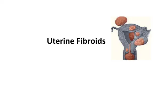
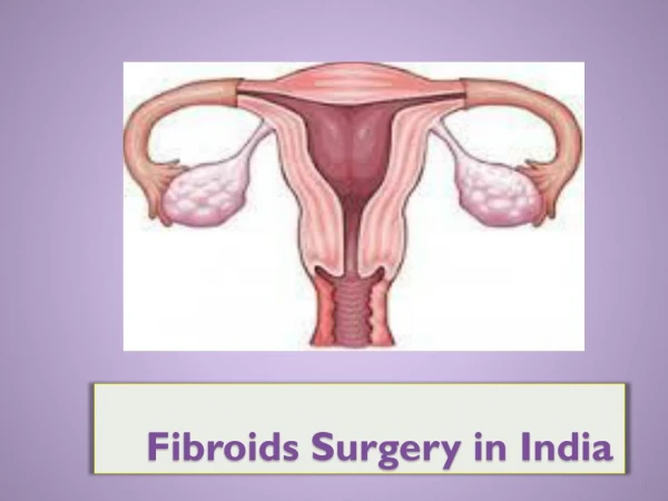
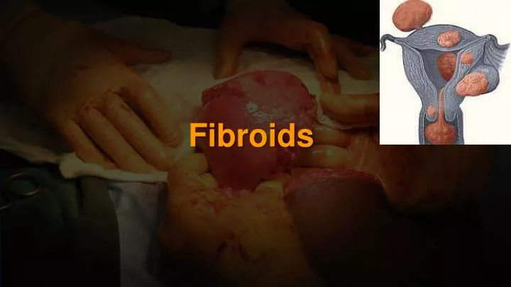
Fibroid Types : Uterine Subserous – Sessile pedunculated Intramural Submucous - Sessile pedunculated Cervical : Anterior, posterior lateral or central Intraligamentous Parasitic
Fibroid Submucous fibroids are classified by European society for gynec endoscopy ( ESGE ): Type 0 – No intramural extension Type I – Intramural extension < 50 % Type II – Intramural extension > 50 %
Fibroid Pathology • Multiple, discrete, spherical, pinkish white, firm capsulated masses . Pseudo capsule is made up of compressed myometrium & areolar tissue. • Microscopically nonstriated muscle fibres are arranged in interlacing bundles of varying size & running in different directions ( whorled appearance ) Varying amount of connective tissue is intermixed with smooth muscle fibres.
Fibroid Pathological variants • Microscopic variants Cellular myoma, mitotically active myoma, bizarre myoma, lipoleiomyoma, • Intravenous leiomyomatosis • LPD – leiomyomatosis peritonealis dissemination • Leiomyosarcoma
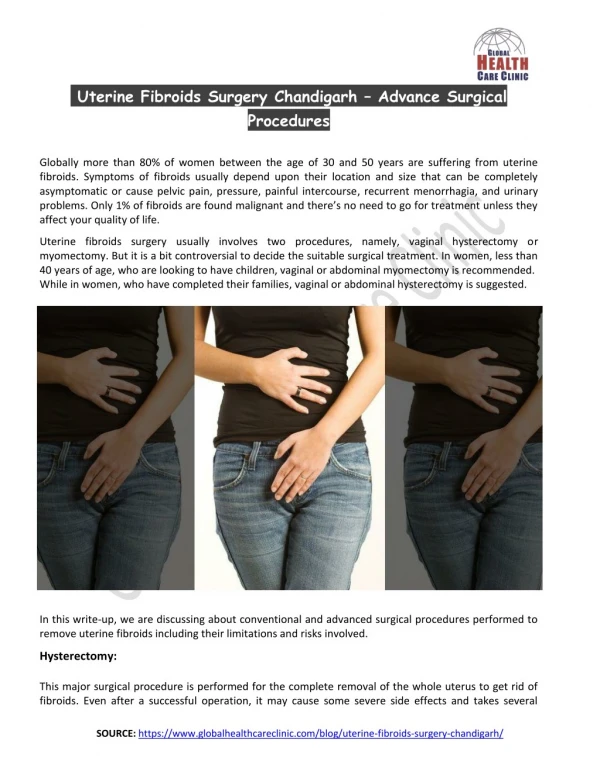
Fibroid Symptoms Asymptomatic - Abnormal uterine bleeding – 30-50% of patients . It is due to ↑↑ surface area, ↑↑vascularity, endometrial hyperplasia, venous obstruction, interference with contractions . - Anemia due to excessive blood loss - Dysmenorrhoea – Spasmodic as well as congestive
Fibroid Symptoms • pelvic pain in 1/3rd patients, backache. • Acute pain due to torsion, infection, expulsion, red degeneration, vascular complication - Pressure symptoms : - Lump in abdomen • Infertility – 2 to 10 % cases * Rare symptoms : Ascites, polycythemia,
Effects of fibroid on pregnancy : • Pregnancy : Abortion Pressure symptoms Malpresentation Retrodisplacement of uterus • Labour : Preterm labour Malpresentation Uterine inertia PPH Dystocia MRP • Puerperium : Subinvolution Sec. PPH Puerperal sepsis Inversion
Effects of fibroid on pregnancy : • Increase in size & softening occurs . Increase occurs mainly in the 1st trimester & in 22 to 32 % cases. • Red degeneration in 2nd trimester – due to rapid growth there is congestion with interstitial hemorrhage & venous thrombosis • Impaction in pelvis • Torsion • Infection • Expulsion • Injury- Pressure necrosis during delivery • Rupture of subserous vein Internal hemorrhage
Fibroid Signs G/E – Anemia due to prolonged heavy bleeding . P/A – If > 12 weeks size , firm, nodular, arising from pelvis, lower limit can’t be reached, relatively well defined, mobile from side to side, nontender, dull on percussion, no free fluid in abdomen P/S – Cervix pulled higher up P/V – Uterus enlarged, nodular. D/D from ovarian tumour Uterus not separately felt , transmitted movement present, notch not felt. P/R – May help in difficult cases .
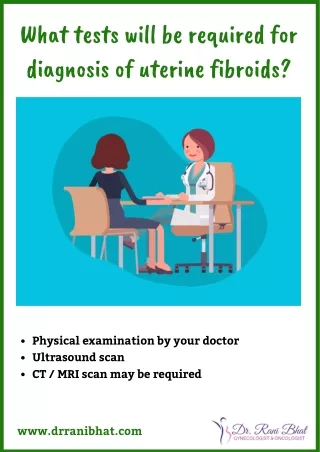
Fibroid Diagnosis • Clinical : From symptoms & signs • USG : Well defined hypoechoic lesions. Peripheral calcification with distal shadowing in old fibroids Adenomyosis is differentiated by diffuse lesion, less echodense , disordered echogenicity & more prominent at or just after menstruation • Hysteroscopy : Submucous fibroids
Fibroid Diagnosis MRI : Most accurate imaging modality for diagnosis of fibroid. It does precise fibroid mapping & characterization Detects all fibroids accurately D/D from adenomyosis D/D from adnexal pathology Ovaries are easily seen Detects small myomas(0.5 cm) H S G : Not done for diagnosis , Done for infertility evaluation filling defects may be seen.
Fibroid D/D • Pregnancy • Adenomyosis • Ovarian tumour • Ectopic pregnancy • Endometriosis • T O mass
Fibroid Pathology Secondary changes :- • Benign : Atrophy, hyaline, necrosis, cystic, calcification,red degeneration, myxomatous ( fatty) , infection • Malignant : Leiomyosarcoma < 1 % in < 50 years < 2 % in > 50 years age
Fibroid Management Expectant : asymptomatic , Size < 12 weeks, near menopause . • Regular follow up every 6 months • Recent guidelines suggest upto 16 wks size however difficult to practice
Medical Management • Not a definitive Rx • For symptomatic relief • Preoperatively to decrease the size • Progestogens, antiprogestogens ( Miefpristone ) androgens ( Danazol, Gestrinone ) & GnRH analogues are used
GnRH analogues Agonists are commonly used drugs :- • Triptorelin ( Decapeptyl) 3.75 mg or leuprolide depot 3.75 mg I/M or Goseraline ( Zoladex) 3.6 mg SC for 3 months • Advantages : Decrease in size of myoma by 20 to 50 % Decrease in bleeding increases Hb level Decreases blood loss during surgery Converts hysterectomy into myomectomy Converts Abd. hyst into vag. hysterectomy Makes hysterectomic resection possible
GnRH analogues • Disadvantages : High cost Hypoestrogenic side effects Effect is reversible Rarely ↑↑ bleeding due to degeneration Occasionally difficulty in enucleation • Antagonist Cetrorelix is used 60 mg I/M repeated after 3-4 months if necessary Initial flare up does not occur
Medical - Newer Therapy SERM – Raloxifen • 60 mg /day is tried for 6 to 12 mths. • Higher doses ( 180 mg) are required for effective decrease in size. • Better if combined with GnRH analogs
Medical - Newer Therapy SPRM - Asoprisnil • 5 to 25 mg/day is used • Mechanism of inhibitory action is not known • Possible risk of endometrial hyperplasia is not studied
Medical - Newer Therapy Mifepristone • 5 – 10 mg is tried • No loss of bone density • Promising results Steinaure et al reviewed 6 trials • Decrease in myoma volume by 26-74 %. • No effect on bone density • Endometrial hyperplasia may limit its longterm use.
Medical - Newer Therapy Aromatase inhibitors • Directly inhibit estrogen synthesis & rapidly produce hypoestrogenic state. Fadrozole is tried in couple of studies • 71 % reduction occurred in 8 weeks • Appears to be promising therapy.
Surgical Management * Hysterectomy Abdominal Vaginal LAVH, TLH * Myomectomy Abdominal Vaginal Hysteroscopic Laproscopic
Surgical Management Vaginal hysterectomy is favoured in following if • Uterus < 16 wks, preferably < 14 wks • No associated pathology like endometriosis , PID, adhesions • Uterus mobile & adequate lateral space in pelvis • Experienced vaginal surgeon
Surgical Management Myomectomy is done in following :- • Infertility • Recurrent pregnancy loss & no other cause • Young patients • Patients who wish to preserve their uterus
Hysteroscopic myomectomy • For submucous myoma causing infertility, RPL, AUB or pain • Criteria :- < 5 cm in size < 50 % intramural component < 12 cm2 uterine size • Gn RH analogue may be given preoperatively • Suspicion of malignancy, infection & excessive mural component contraindicates surgery • Advantages are short procedure , rapid recovery & all disadvantages of laprotomy avoided.
Laproscopic myomectomy In 3 phases excision of myoma, repair of myometrium & extraction • Suitable for subserous & intramural fibroids upto 10 cm size • Complications are those of operative laproscopy + myomectomy • Fibroid excised are remoyed by electronic morcellators or through posterior colpotomy incision vaginally.
Abdominal myomectomy - Other factors for infertility should be ruled out - Consent for hysterectomy - Blood ‘X’ matched & ready - Pap’s smear & endometrial sampling to rule out malignancy - Medical or mechanical means to control blood loss Bonney’s Myomectomy clamp, rubber tourniquet, manual ( finger compression) pressure at isthmic region or use of vasopressin 10 – 20 units diluted in 100ml saline infiltrated before putting the incision .
Abdominal myomectomy • Minimum incisions are kept – preferably single midline vertical, lower, anterior wall . • Removal of as many fibroids as possible through one incision & secondary tunnelling incisions. • Meticulous closure of all dead space. • Proper haemostasis • Multiple small fibroids can be removed enbloc by wedge resection. • Measures for adhesion prvention should be taken.
Abdominal myomectomy • Morcellation – Deeply embedded tumours are best removed by cutting them into bits. • Bonney’s hood – for posterior fundal large fibroid transverse fundal incision posterior to tubal insertion is made & uterine wall after enucleation is sutured anteriorly covering the fundus as a hood. • Complications of myomectomy like hemorrhage & infection are less in modern times.
Vaginal myomectomy • Submucous pedunculated or small sessile cervical fibroids are removed vaginally. • Ligation of pedicle if accessible • Twisting off the fibroids if pedicle not accessible in case of small & medium size fibroids • To gain access to pedicle of higher & big fibroid incision on the cervix can be made.
Surgical Management Laproscopic myolysis :- • By ND-YAG laser or long bipolar needle electrode thro. Laproscope blood supply of myoma is coagulated. • Without blood supply myoma atrophies. • Applicable to 3 -10 cm size & myomas < 4 in number * Cryomyolysis is under investigation
Uterine artery embolization • By interventional radiologist • Catheter is passed retrograde thro. Right femoral artery to bifurcation of aorta & then negotiated down to opposite uterine artery first. • Polyvinyl alcohol ( PVA ) particles ( 500-700 um) or gelfoam are used for embolization. • 60 – 65 % reduction in size of fibroid • 80 – 90 % have improvements in menorrhagia & pressure symptoms
Uterine artery embolization • High vascularity & solitary fibroid are associated with greater chance of longterm success. • Pregnancy, active infection & suspicion of malignancy are absolute C I . • Desire for fertility is also a contraindication to UAI • The risk of ovarian failure must be counselled • Post embolization syndrome ( fever ,vomiting, pain) can occur
Fibroid Newer Management Mirena : • Third generation IUCD • Contains Progesteron LNG 60 mg releasing 20 ug /day • Fibroids decreases in size 6 – 12 mths of use. • May have variable effects on uterine myomas depending upon balance of growth factors • Couple of studies have shown beneficial results • Suitable for those who also desire contraception
Newer Management- MRGFUS • Permitted by FDA since 2004 • MRI guidance is used to direct ultrasound to tissues to elicit coagulative necrosis via thermal ablation.
Newer Management- MRGFUS • Fasting overnight • Shaving of lower abdomen • Foley’s catheter • Sonications of 20 to 40 seconds interval with 80 – 90 seconds cooling
Research Lanreotide a long acting somatostatin analog reduces GH secretion 30 mg depot reduced fibroid size by 41.6 % Targetting growth factors that are involved in angiogenesis or fibrosis Pirfenidone an antifibrotic agent is under trial