Download
1 / 19
210 likes | 652 Views
Bones: Tissue and Organ. Bones can be referred to as either a tissue ( osseous ) or an organ Bone referred to as a connective tissue consists of: cells extracellular matrix (ground substance + fibers) this living/dynamic tissue is capable of growth and repair

E N D
Bones: Tissue and Organ • Bones can be referred to as either a tissue (osseous) or an organ • Bone referred to as a connective tissue consists of: • cells • extracellular matrix (ground substance + fibers) • this living/dynamic tissue is capable of growth and repair • Bone referred to as an organ (particular bones of the body) consists of: • osseous tissue • nervous tissue • epithelial and muscle tissue (blood vessels) • Ex. femur, humerus, clavicle, sternum…
Function of Bones • Support • form the framework that supports the body • Protection • provide a protective cavity for the brain (skull), spinal cord (vertebrae), and vital organs (rib cage) • Movement • provide attachment points for skeletal muscles and are used as levers • Mineral storage • reservoir for calcium and phosphorus • Blood cell formation (hemopoiesis) • occurs within the red bone marrow of bones
Gross Anatomy of Bones: Bone Textures • Compact bone • dense and heavy bone • Spongy bone • porous and light bone • formed by rods and plates of bone called trabeculae • spaces filled with red bone marrow • provides strength with little weight • The bones of the body contain both bone textures • the outer layer is made of compact bone • the inner portion of bones is made of spongy bone
Structure of Long Bones • Diaphysis • tubular shaft of compact bone that surrounds the medullary cavity • during growth periods contains red bone marrow • in adults, contains yellow bone marrow (fat) • Epiphyses • expanded ends of long bones • exterior is compact bone, and the interior is spongy bone • superficial surface covered in articular (joint) cartilage • epiphyseal plate (line) separates the diaphysis from the epiphyses and represents location of bone lengthening (growth)
Cells of Osseous Tissue • Osteoblasts • found in periosteum and endosteum • cells that make (deposit) the matrix of osseous tissue • increase bone density • Osteoclasts • found in periosteum and endosteum • cells that break down (resorb) the matrix of osseous tissue • decrease bone density • Osteocytes • cells that are surrounded by matrix (located in a lacunae) that are not depositing or resorbing matrix
Composition of Bone (Extracellular) Matrix • Inorganic components • hydroxyapatites • calcium and phosphate deposits • 65% of bone by mass • responsible for bone hardness • Fibers • collagen • provides an ability of bone to flex slightly without fracturing
Bone Histology • The basic structural unit of bone is called the osteon (haversian system) • a central canal holding blood vessels and nerves that are parallel to the longest dimension of the bone is surrounded concentric cylinders of bone called lamellae • osteocytes found between lamellae are connected to each other by tiny cracks in the lamellae called canaliculi • Perforating canals • canals that are perpendicular to the central canals joining them to the blood and nerve supply at the superficial surface of the bone
Coverings of Bone • There are 2 surfaces of a bone that are covered with a thin layer of connective tissue • the most superficial surface is covered by the periosteum • the internal surface of the central and perforating canals is covered with endosteum • Both the periosteum and the endosteum contain osteoblasts and osteoclasts which are capable of increasing and decreasing the amount of bone tissue • bone remodeling
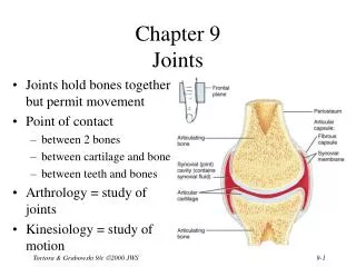
Joints (Articulations) • Site where two or more bones meet • Weakest parts of the skeleton • Functions include: • provide mobility for the skeleton • bones move in relation to one another where the joints serve as a fulcrum (pivot point) and the associated bones serve as levers • hold the skeleton together
Classification of Joints • The three functional classes of joints are: • Synarthroses • immovable • Amphiarthroses • slightly movable • Diarthroses • freely movable • The three structural classifications are: • Synovial • Fibrous • Cartilaginous
Synovial Joints • Those joints in which the articulating bones are separated by a joint capsule filled with synovial fluid • all are freely movable (diarthroses) • found mainly in arm and legs
Cartilaginous Joints • Articulating bones are united by cartilage • most are slightly movable (amphiarthroses) • include intervertebral discs
Fibrous Joints • The bones are joined by dense connective tissue containing a high amount of collagen • most are immovable (synarthroses) • include the bones of the skull