Download
1 / 64
670 likes | 967 Views
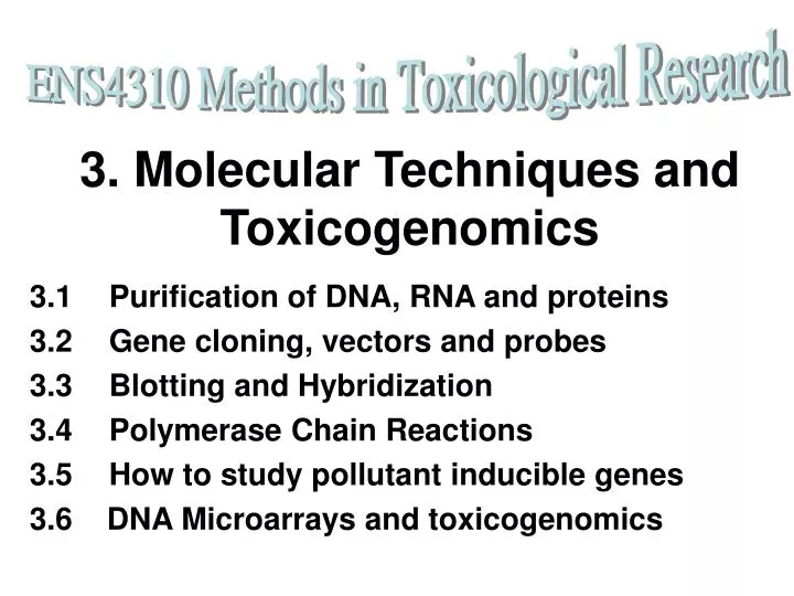
ENS4310 Methods in Toxicological Research. 3. Molecular Techniques and Toxicogenomics. 3.1 Purification of DNA, RNA and proteins 3.2 Gene cloning, vectors and probes 3.3 Blotting and Hybridization 3.4 Polymerase Chain Reactions 3.5 How to study pollutant inducible genes

E N D
ENS4310 Methods in Toxicological Research 3. Molecular Techniques and Toxicogenomics 3.1 Purification of DNA, RNA and proteins 3.2 Gene cloning, vectors and probes 3.3 Blotting and Hybridization 3.4 Polymerase Chain Reactions 3.5 How to study pollutant inducible genes 3.6 DNA Microarrays and toxicogenomics
3.1. Purification of genomic DNA • Preparation of nuclei from white blood cells, i.e. blood samples. • Any tissues, such as muscle/flesh and testis are excellent tissues for genomic DNA preparation. • Liquid Nitrogen grounding method (traditional method). • Digestion with proteinase K in buffer with EGTA and EDTA, which are DNase inhibitors. • Phenol-chloroform extractions followed by ethanol precipitation, or dialysis. • Replaced by Silica-based DNA purification method.
Apoptotic DNA analyzed on agarose gel electrophoresis after ethidium bromide staining and visualized on UV light. - + DNA ladder formed in apoptotic DNA. Why ladder was formed?
3.1.2 Purification of RNA • Total RNAs include messengerRNA (mRNA), ribosomal RNA (rRNA), and transfer RNA (tRNA). • Avoid and degrade ribonuclease, use guanidine thiocyanate or guanidine HCl as protein denaturant. • Phenol-chloroform extractions to remove protein and nuclease, finally ethanol precipitation of nucleic acids. • Only RNA is able to get into aqueous phase, degraded protein trapped in the interphase. • Use oligo-dT cellulose to capture polyadenylated mRNA in high salt, elute in water and use SDS as detergent and ribonuclease inhibitor.
Purification of polyadenylated messenger RNAs Adapted from Molecular Cell Biology, 2000.
Total RNA samples resolved on denaturing agarose gel with formamide buffer and 2.2 M formaldehyde. - kb 9.4- 7.4- 4.4- 28s rRNA 2.4- 1.4- 18s rRNA 0.4- 5s RNA +
3.1.3 Purification of proteins • Identify source of the proteins of interest: microsome, cytosol, nucleus, membrane. • Use centrifugation methods in buffer with protease inhibitors. • Precipitation in salt or acetone. • Sizes fractionation on columns. • Separate according to charges on ion-exchange column. • Separate according to hydrophobicity on reverse phase High Pressure Liquid Chromatography.
Sub-cellular Fractionation • Use of buffer (isotonic) • Use of detergents, SDS, Triton, NP40 (ionic and non-ionic detergents) • Use of centrifugation, e.g. Ultra-centrifugation (100,000 xg) • Salt precipitations or acetone precipitation at the end
Centrifugations to Isolate Organelles Homogenization in buffer on ice centrifuge at 4oC, 1,500 x g for 10 min By high speed centrifuge pellet (nuclei + membrane) supernatant centrifuge at 4oC, 9,000 x g for 30 min dissolved in 7 ml of homogenizing buffer supernatant (post-mitochondrial fraction) pellet, dissolved in 7ml homogenizing buffer (mitochondrial fraction) carefully added on cushion of 4oC pre-chilled homogenizing buffer with 2M sucrose centrifuge at 4oC, 105,000 x g for 60 min ultracentrifuge centrifuge at 4oC, 70,000 x g for 30 min supernatant (cytosolic fraction) pellet, dissolved in 7 ml homogenizing buffer (microsomal fraction) pellet, dissolved in 7 ml homogenizing buffer (nuclear fraction) supernatant (plasma membrane) Adapted from David L. Spector, et al., “Cells – A Laboratory Manual”, vol.1, Cold Spring Harbor Laboratory Press, chapter 34, 43
3.2 Gene cloning, vectors and probes 3.2.1 Gene hunting 3.2.2 Cloning vectors: plasmid and phage 3.2.3 Restriction enzymes 3.2.4 Genomic DNA libraries 3.2.5 cDNA libraries 3.2.6 Colony lifting and library screening
3.2.1 Gene Hunting • Functional cloning and positional cloning • Reverse genetics: purify protein first, get partial amino acid sequences, design primers as probe or for PCR, screen cDNA libraries. (Functional cloning) • Positional cloning: use genetic markers to localize chromosomal region for a particular gene mutation or defects from single gene.
Reverse Genetics: From Protein to Gene • Isolate protein • Partial amino acid sequence (e.g. AhR cloning) • Predict mRNA sequence to design oligonucleotide sequences • Use oligonucleotides as probe or primers for PCR • Screen cDNA library to isolate cDNA encoding for the protein. • Alternatively use RT-PCR (reverse transcription-polymerase chain reaction) • Use cDNA clone as probe to isolate gene.
3.2.2 Cloning vectors: plasmid and phage • Gene cloning with insertion of DNA into a vector which could be introduced into a host for propagation or mass production. • Use restriction enzymes to cut and past DNA pieces. • Vector DNA must have three components: origin of replication, antibiotic resistant gene, marker gene with multiple cloning sites. • Plasmid is extra-chromosomal circular DNA. • Phage is linear DNA to be packaged in phage particles. • Other vectors include phagemid, cosmid, yeast artificial chromosome, etc.
Three Major components in a plasmid vector: 1. Origin of replication 2. Antibiotic resistant gene 3. Multiple cloning site (in marker gene’s coding region) Adapted from Molecular Cell Biology, 2000.
Different cloning vectors for genomic library construction • Plasmid: 0 to 10 kbp inserts, suitable only for bacterial genomic libraries. (why? Think?) • Phage: 9-22 kbp, suitable for organisms with a larger genome. • Cosmid: 25-50 kbp, suitable for genes with larger sizes. • YAC (yeast artificial chromosome): 50-100 kbp or even 1000 kbp, suitable for chromosome jumping or walking.
3.2.6 Colony lifting and library screening • Plate out library onto agar plate with top agarose of around 30,000 to 50,000 plaques or colonies per plate. • You need 3 times more clones to be plated and spread out if you want to get one clone by chance. • In other words, if you want to get a single gene (say 15kb, 1.5 x 10³bp) from a genome size of 1N say 1.5 X 10⁹ bp, you need to spread out at least 3 times of 106 colonies, that is 3 X 10⁶ colonies at one time. That means you need to plate out 60 plates of 50,000 colonies each.
1. Principle of colony lifting of bacterial cells onto nylon membrane, 2. Treated with NaOH to get DNA onto the nylon membrane, 3. Perform hybridization to identify locations of colony that give signals!! Adapted from Molecular Cell Biology, 2000.
Hybridization of labeled DNA probe to DNA fixed on nylon membrane lifted from agar plates. Adapted from Molecular Cell Biology, 2000.
Screening of expression library Antibodies can also be used to screen an expression library with colonies showing the expressed protein after induction by IPTG. Adapted from Molecular Cell Biology, 2000.
Revision Questions for Recombinant DNA methods • Compare and contrast the methods use for the construction of cDNA library and genomic library. • Compare and contrast different vectors used for cloning. What are essential elements in a vector? • Why do we want cDNA clones and what can we use cDNA clones for? (Use Ah receptor as an example, can be expressed and used for binding assay) • How about cloned gene promoter? Cloned genes are for further biotech manipulations, see following example, gene promoter could be used as detection system in recombinant cell-line.
3.3 Blotting and Hybridization • Transfer of biomolecules to a solid and thin support such as nylon membrane. • Southern: transfer of DNA from agarose gel. • Northern: transfer of RNA from agarose gel. • Western: transfer of protein from polyacrylamide gel. • Transfer could be done via capillary action, vacuum, and electrical mean (western only). • Cloned genes or PCR products could be used as probes for Northern and Southern blot analyses. Western blot need antibodies to detect.
3.3.1 Southern Blotting Capillary Action OR Vacuum Blotting High salt buffer to transfer DNA to a solid (soft) support of nylon membrane.
3.3.2 Northern Blot Analysis: RNA study Watson, 1994.
3.3.3 Electro Blotting Western Blotting for Protein Analyses Semi-Dry Transfer: from SDS-PAGE to nylon membrane
Can detect protein sizes Watson, 1994. 2.3.4. Western Blot Analysis
3.4 Polymerase Chain Reactions • The PCR is a simple method to make copies of a particular DNA fragment using a thermo- (heat) stable DNA polymerase. • It consists of cycles of denaturation of template DNA at 94-95 C, annealing with a pair of gene specific primers, and extension of strands with a heat stable (thermostable) DNA polymerase.
3.4.1. Principle of Polymerase Chain Reaction
PCR • Single strand DNA primer pair is designed from the two ends of the DNA fragment that you want to amplify. • Following PCR cycles, million copies of DNA fragments could be produced from a single copy of any DNA or gene. • Drs Kary Mullis and Michael Smith were awarded 93 Nobel in Chemistry for their invention of PCR and chemical synthesis of DNA oligonucleotide respectively.
3.4.2. 1st strand cDNA synthesis Reverse Transcription-PCR RT-PCR
RT-PCR analysis of hepatic RNA samples from saline control (lane 2) and cadmium treated tilapia using metallothionein gene specific primers. Lane 4 is negative control without cDNA sample, lane 1 and 5 are DNA size marker. Agarose (2%) gel was stained with ethidium bromide after electrophoresis and visualized on UV light transilluminator.)
3.4.3 Other applications of PCR • Amplify any gene fragment with known sequence. • Perform quantifiable real time PCR. • DNA sequencing. • Detection of gene mutation with real time PCR or DNA sequencing to look at site specific single mutation. • DNA finger-printing: short primers to obtain different patterns or obtain variable gene regions. • Detection of environmental pathogens. • Detection of DNA sequence for forensic applications.
3.4.4. Real-time PCR • What affect the appearance of amplicons? Ans: amount of template, primer matching, annealing temperature, PCR cycle number. • Laser detection of fluorescent dye labeled amplicon over time from 2-40 cycles • Compare slopes to evaluate the amount of amplicon produced in the PCR reaction • Taq-Man detection of specific amplicon or SYBR green detection methods • Quantitative method, RNA standard can be used.
Detection chemistries • Four common ways: • DNA binding dyes • E.g. SYBR Green • Hydrolysis probes • TaqMan • Hybridisation probes • E.g. Light cycler • Hairpin probes • Molecular beacons 3. PCR Quantification
TaqMan Probe is Gene specific and can provide accurate detection of target gene’s mRNA level by using RT-PCR Real Time PCR using Gene Specific TaqMan Probe (Fluorogenic 5’ Nuclease Chemistry: reporter and quencher stay together give no fluorescent signal, however, the reporter dye shows fluorescence when detached from quencher)
2. Theory of Real-time PCR • CT - threshold cycle: • the first significant increase in the amount of PCR product correlates to the initial amount of target template • CTrepresents the starting copy no. in the original template • Early exponential phase: • PCR is just began • The amount of fluorescence has reached a threshold where it is significantly higher than background (usually 10 times the standard deviation of the baseline) • Linear ground phase: • PCR is just began • Fluorescence emission at each cycle has not yet risen above background • Baseline fluorescence is calculated at this time PCR can be broken into 4 major phases
2. Theory of Real-time PCR • Log-linear phase: • PCR reaches its optimal amplification period with the PCR doubling after each cycle in ideal reaction conditions • Plateau phase: • The plateau stage is reached when reaction components become limited and the fluorescence intensity is no longer useful for data calculation
Fluorescent signals are detected with laser detection system. We may compare the Ct values (threshold) All detection chemistry give similar results, the question is how do you want to quantify the data.
Validation experiment for comparative CT method ABI-7700 User Bulletin #2 GAPDH is for normalization, serving as a control in this experiment. 3. PCR Quantification
Comparative CT method-I ABI-7700 User Bulletin #2 3. PCR Quantification
Comparative CT method - II ABI-7700 User Bulletin #2 3. PCR Quantification
Q PCR Quantification control D Ct = target - ref ref control D Ct = 9.70 target control av =19.93 av =29.63 D Ct = target - ref experiment targettreated D Ct = -1.7 reftreated Difference = DCt-DCt = DDCt = (-1.7) -9.70 = -11.40 av =18.03 av =19.80 Exercise: By 2 –∆∆CT, fold change=??? 2702
3.5 How to study pollutant inducible genes? • Differential screening • Subtractive hybridization • Differential display by RT-PCR • DNA microarray method (can study both inducible and depressed genes)
3.5.1 Differential screening
Example: Identification of dioxin inducible genes • CYP1A1, 1A2 • ALDH-3 (aldehydehrogenase) • UGT1A1 • GSTYa • Plasminogen activator inhibitor, PAI-2 • Interleukin-1β Drug metabolizing enzymes, growth and differentiation controller, and related to Immune System
3.5.3 Differential Display DD RT-PCR is a powerful tool in searching for inducible genes or depressed genes. Same primers are used to amplify cDNAs from different samples (treated and untreated)