Download
1 / 74
890 likes | 2.56k Views
Diagnosis and Management of Acute Stroke. Briana Witherspoon DNP, ACNP-BC. Stroke Objectives. Review etiology of strokes Identify likely location/type of stroke based of physical exam Acute management of ischemic stroke Acute management of hemorrhagic stroke . Stroke Fast Facts .

E N D
Diagnosis and Management of Acute Stroke Briana Witherspoon DNP, ACNP-BC
Stroke Objectives • Review etiology of strokes • Identify likely location/type of stroke based of physical exam • Acute management of ischemic stroke • Acute management of hemorrhagic stroke
Stroke Fast Facts • Affects ~ 800, 000 people per year • Leading cause of disability, cognitive impairment, and death in the United States • Accounts for 1.7% of national health expenditures. • Estimated U.S. cost for 2012 = $71.5 billion • Mostly hospital (esp. LOS) & post stroke costs • Appropriate use of IV t-PA s long-term cost • Appropriate billing for AIS w/ thrombolysis ( hospital reimbursement from $5k to $11.5k) Stroke. 2013;44:2361-2375
Where We’re Headed • By 2030 ~ 4% of the US population over the age of 18 is projected to have had a stroke • Between 2012 and 2030, total direct stroke-related medical costs are expected to increase from $71.55 billion to $183.13 billion • Total annual costs of stroke are projected to increase to $240.67 billion by 2030, an increase of 129% Stroke. 2013;44:2361-2375
Intracerebral Hemorrhage Bleeding into brain 10% Subarachnoid Hemorrhage Bleeding around brain 5% Ischemic Stroke Clot occluding artery 85% Three Stroke Types www.acponline.org/about_acp/chapters/ok/gordon.ppt
http://www.phillystroke.org/content/learn_about_stroke/act_fast.asphttp://www.phillystroke.org/content/learn_about_stroke/act_fast.asp
NIHSS • NIHSS (National Institute of Health Stroke Scale) • Standardized method used by health care professionals to measure the level of impairment caused by a stroke • Purpose • Main use is as a clinical assessment tool to determine whether the degree of disability is severe enough to warrant the use of tPA • Another important use of the NIHSS is in research, where it allows for the objective comparison of efficacy across different stroke treatments and rehabilitation interventions • Scores are totaled to determine level of severity • Can also serve as a tool to determine if a change in exam has occurred
Breaking Down the Scale • 13 item scoring system, 7 minute exam • Integrates neurologic exam components • CN (visual), motor, sensory, cerebellar, inattention, language, LOC • Maximum score is 42, signifying severe stroke • Minimum score is 0, a normal exam • Scores greater than 15-20 are more severe
NIHSS cont. • NIHSS Interpretation
NIHSS and Outcome Prediction • NIHSS below 12-14 will have an 80% good or excellent outcome • NIHSS above 20-26 will have less than a 20% good or excellent outcome • Lacunar infarct patients had the best outcomes Adams HP Neurology 1999;53:126-131 Baseline NIH Stroke Scale score strongly predicts outcome after stroke (TOAST)
Etiology of Ischemic Strokes LARGE VESSEL THROMBOTIC: Virchow’s Triad…. • Blood vessel injury • HTN, Atherosclerosis, Vasculitis • Stasis/turbulent blood flow • Atherosclerosis, A. fib., Valve disorders • Hypercoagulable state • Increased number of platelets • Deficiency of anti-coagulation factors • Presence of pro-coagulation factors • Cancer
Etiology Of Ischemic Stroke: LARGE VESSEL EMBOLIC: • The Heart • Valve diseases, A. Fib, Dilated cardiomyopathy, Myxoma • Arterial Circulation (artery to artery emboli) • Atherosclerosis of carotid, Arterial dissection, Vasculitis • The Venous Circulation • PFO w/R to L shunt, Emboli
Determining the Location • Large Vessel: • Look for cortical signs • Small Vessel: • No cortical signs on exam • Posterior Circulation: • Crossed signs • Cranial nerve findings • Watershed: • Look at watershed and borderzone areas • Hypo-perfusion
Cortical Signs • If present, think LARGE VESSEL stroke
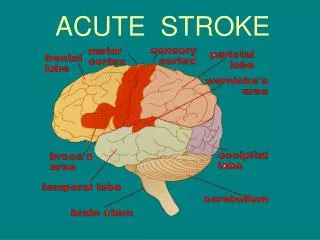
Large Vessel Stroke Syndromes • MCA: • Arm>leg weakness • LMCA cognitive: Aphasia • RMCA cognitive: Neglect,, topographical difficulty, apraxia, constructional impairment • ACA: • Leg>arm weakness, grasp • Cognitive: muteness, perseveration, abulia, disinhibition • PCA: • Hemianopia • Cognitive: memory loss/confusion, alexia • Cerebellum: • Ipsilateral ataxia
Aphasia • Broca’s • Expressive aphasia • Left posterior inferior frontal gyrus • Wernicke’s • Receptive aphasia • Posterior part of the superior temporal gyrus • Located on the dominant side (left) of the brain
Case 1 • 74 year old African American female with sudden onset of left-sided weakness • She was at church when she noted left facial droop • History of HTN and atrial fibrillation • Meds: Losartan
Case 1 • BP- 172/89, P– 104, T- 98.0, RR– 22, O2- 94% • General exam: Unremarkable except irregular rate and rhythm • NEURO EXAM: - Speech dysarthric but language intact - Right gaze preference - Left facial droop - Left- sided hemiplegia - Neglect
Case 1 • Right MCA infarct, most likely cardioembolic from atrial fibrillation • Patient underwent mechanical thrombectomy with intra-arterial verapamil, clot removal successful • Excellent recovery – patient was discharged 48 hours later on Coumadin
Determining the Location • Large Vessel: • Look for cortical signs • Small Vessel: • No cortical signs on exam • Posterior Circulation: • Crossed signs • Cranial nerve findings • Watershed: • Look for watershed pattern • S/S of Hypo-perfusion
Etiology of Stroke SMALL VESSEL (Lacunes <1.5cm) • Risk Factors • HTN • HLD • DM • Tobacco Use • Sleep apnea
Case 2 • 85 year old male who woke up with left face, arm, and leg numbness • History of HTN, DM, and tobacco use • Meds: Insulin, aspirin
Case 2 • BP- 168/96, P– 92 • General exam: Unremarkable, RRR • NEURO EXAM: - Decreased sensation on left face, arm, and leg
Case 2 • Right thalamic lacunar infarct • Not a candidate for intervention (WHY?) • Discharged to rehab 72 hours after admission
Determining the Location • Large Vessel: • Look for cortical signs • Small Vessel: • No cortical signs on exam • Posterior Circulation: • Crossed signs • Cranial nerve findings • Watershed: • Look at watershed and borderzone areas • Hypo-perfusion
Brainstem Stroke Syndromes • Rarely presents with an isolated symptom • Usually a combination of cranial nerve abnormalities, and crossed motor/sensory findings such as: • Double vision • Facial numbness and/or weakness • Slurred speech • Difficulty swallowing • Ataxia • Vertigo • Nausea and vomiting • Hoarseness
Case 3 • 55 year old male with acute onset of right sided numbness and tingling, left sided face pain and numbness, gait imbalance, nausea/vomiting, vertigo, swallowing difficulties, and hoarse speech • History of CAD s/p CABG, DM2, HTN, HLD, OSA • Meds: Aspirin, plavix, insulin, lipitor, metoprolol, lisinopril
Case 3 • NEURO EXAM: BP- 194/102, P– 105 • General exam: Unremarkable, RRR • NEURO EXAM: - Decreased sensation on left face - Decreased sensation on right body - Left ataxia on FNF, and unsteady gait - Voice hoarse - Nystagmus
Case 3 • Brainstem Stroke • Received IV tPa • Post-tPa symptoms greatly improved regained sensation, ataxia resolved • Discharged home with out patient PT/OT
Determining the Location • Large Vessel: • Look for cortical signs • Small Vessel: • No cortical signs on exam • Posterior Circulation: • Crossed signs • Cranial nerve findings • Watershed: • Look for the watershed pattern • Think about reasons of hypo-perfusion • Hypotension • Stenosed vessel, etc
Case 4 • 56 year old female who upon waking post-op after elective surgery was found to have L sided weakness and neglect • History of HTN • Meds - Lisinopril
Case 4 • BP- 132/74, P– 84 • General exam: Unremarkable, RRR • NEURO EXAM: - Left face, arm, and leg weakness - Neglect - DTR’s brisk on the left, toe up on left
Case 4 • Right hemisphere watershed infarct secondary to hypoperfusion in the setting of Right ICA stenosis • On review of anesthesia records, blood pressure dropped to 82/54 during the procedure • Patient was discharged to in-patient rehab
Etiology of ICH • Traumatic • Spontaneous • Hypertensive • Amyloid angiopathy • Aneurysmal rupture • Arteriovenous malformation rupture • Bleeding into tumor • Cocaine and amphetamine use
Causes of ICH http://spinwarp.ucsd.edu/neuroweb/Text/non-trauma-ER.htm