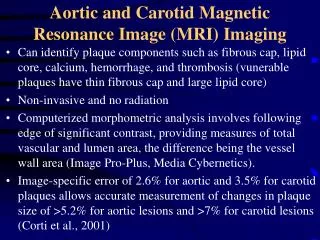
Download
1 / 16
160 likes | 252 Views
Automated versus human in vivo segmentation of Carotid Plaque MRI. C Oppenheim 1 , R van 't Klooster 2 , R Marsico 1 , O Naggara 1 , O Eker 1 , RJ van der Geest 2 , IM Adame 3 , E Touzé 1 , Meder JF 1 . 1. Université Paris Descartes, INSERM U894 Sainte-Anne hospital, Paris, France

E N D
Automated versus human in vivo segmentation of Carotid Plaque MRI C Oppenheim1, R van 't Klooster2, R Marsico1, O Naggara1,O Eker1, RJ van der Geest2, IM Adame3, E Touzé1, Meder JF1. 1. Université Paris Descartes, INSERM U894 Sainte-Anne hospital, Paris, France 2. Division of Image Processing, Department o f Radiology, Leiden University Medical Center, Leiden, The Netherlands 3. Medis medical imaging systems bv, Leiden, The Netherlands
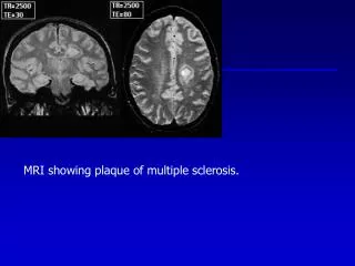
Introduction Beyond the lumen … Hemorrhage Calcium Fibrous Lipid core Initial MRA One year follow-up
Stable Unstable
methods How to study atherosclerotic plaques in vivo ? 1. Clarke, et al. Magn Reson Med. 2003 2. Ronen, et al. Magn Reson Med. 2007 3. Liu, et al. Magn Reson Med. 2006 4. Binjian et al. Magn Reson Med. 2008 5. Adame IM, et al. MAGMA. 2004 6. Karmonik et al.. IEEE Trans Bio. Eng. 2009 7. Kerwin et al. Top Magn Reson Imaging 2007 8. Touzé et al. Stroke 2007
Purpose To compare the detection and quantification of the main MR plaque components in vivo using • manual approach • automatic approach
Material and Methods 60 patients with carotid stenosis • Extracted from a multicentric prospective study (HIRISC) • Asymptomatic high grade stenosis or symptomatic moderate stenosis • With high quality MRI (at least ¾ sequences = excellent) Training set n=20 Study group n=40 No differences for age, sex, degree of stenosis, % symptomatic stenosis
Training set, n=20 Study group, n=40 Supervised classifier Software Q Plaque (Medis medical imaging bv)
Manual versus Automatic méthodes • Study group : 40 patients, 344 sections • Qualitative Analysis: Kappa coefficient (IC 95%) • Quantitative Analysis: • Intraclass coefficient correlation (IC 95%) • Linear correlation between volumes • Bland Altman plots
Results Qualitative analysis (per patient)
Results Quantitative analysis Lipids Hemorrhage Calcium Fibrous
Results LIPIDS: Bland Altman plots Volume (Manual – Automatic) Mean Volume, mm3
Discussion • The automaticanalysis of atherosclerotic components on MRI is possible • Identification : • Kappa at least substantial for all components • Slightlybetter for hemorrhage (highcontrast on all sequences) • Quantification : • Best for hemorrhage, fibrous tissue, lipidcore < 100 mm3 • Poor for Calcium • In patients withoutclearsurgical indications usingdifferent MR plateforms
discussion Limites • High quality images • Results could be improved with a larger training set • Fibrous cap ? • No reference (patients not scheduled for endarteriectomy)
Conclusion : Automatic plaque analysis Day 0 • A gain in time • No variability • Potentially interesting for • Large multicentric therapeutic trials • Longitudinal studies • Could help the integration of HR MRI of atherosclerosis in clinical practice 1 year